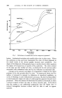
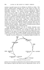
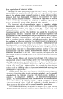
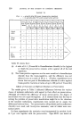
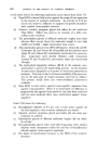
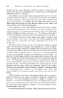
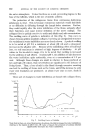
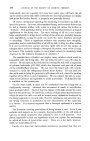
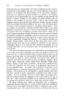
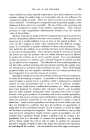
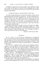
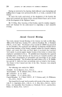
THE JOURNAL OF THE SOCIETY OF COSMETIC CHEMISTS This edition is published by THE SOCIETY OF COSMETIC CHEMISTS OF GREAT BRITAIN Publications Office: 54, Woodlands, London, N.W. 11. VOLUME IX JULY 1958 NUMBER 4 SKIN AND HAIR PIGMENTATION A.L. LORINCZ, M.D.* Read at the Cosmetic Seminar, Chicago, on loth september 1957. The formation and l•rol•erfies of melanin are discussed at some length and there are also references to o•er l•rima• lfigments. L.ARCEL¾ on the basis of studies by reflectance spectrophotometry five primary pigments have been recognized as being responsible for the color of normal human skin.•,", a These are melanin, oxyhemoglobin, reduced hemoglobin, carotene, and melanoid. Figure 1 shows the reflectance spec- trum of normal white human skin and its component pigments. Melanin shows a general marked absorption at the violet end of the visible spectrum with no clear-cut absorption bands oxyhemoglobin has a couple of sharp absorption bands at 542 and 576 m/• reduced hemoglobin has a single broad absorption band at 556 m/• carotene has one at 482 m/• and melanoid has some general absorption at the violet end of the spectrum like melanin but, in addition, has a distinct absorption band at 400 m/•. The physical distribution patterns of these pigments and the thickness and optical properties of the overlying colorless tissue layers, are additional factors that modify the final color effects produced by these pigments. The farther below the skin surface any dark pigment is situated, the bluer it appears, because of the Tyndall light-scattering phenomenon. In this phenomenon, light, in traversing a turbid medium, retains a larger proportion of long or red wave lengths, while the light scattered to the sides and back to the source contains a correspondingly larger proportion of the shorter or •blue wave lengths. This simply means that blue light is more readily scattered by tiny particles than is red light. It is this scattering back of the blue components of light by overlying turbid tissue, that gives the bluish color to blue nevi, Mongolian spots, the black ink of tattoos and to purpuric * Department of Medicine, University of Chicago, U.S.A. 197

198 JOURNAL OF THE SOCIETY OF COSMETIC CHEMISTS 60 50 2O 420 460 500 540 580 620 660 700 WAVELENGTH Fig. 1. Reflectance of normal skin and its component pigments. a lesions. Anatomical variations also modify skin color in other ways. Where the epidermis is thin and more transparent the color of blood pigments in the small vessels of the dermal papillae becomes more prominent. The degree of development of the granular layer is another factor which regulates the degree of transparency or opacity of the epidermis. The water content of the skin can also modify its color, as illustrated by the general pallor associated with oedematous skin and by the whiteness of macerated skin. Changes in the amount and degree of oxygenation of blood in the upper portions of the skin greatly affect its color. If oxygenated blood near the surface is increased in amount by dilatation of superficial capillaries, as occurs in blushing or acute inflammatory processes, the skin becomes pinker or red because the bright red color of oxyhemoglobin pigment becomes more prominent. When the amount of blood near the surface is decreased, as when a person is about to faint, the skin, of course, becomes pale. If the blood near the surface is not sufficiently oxygenated, then the bluish color of reduced hemoglobin becomes evident and the skin is said to appear cyanotic.
Purchased for the exclusive use of nofirst nolast (unknown)
From: SCC Media Library & Resource Center (library.scconline.org)