SKIN AND HAIR PIGMENTATION 199 The yellow pigment, carotene, is present in the subcutaneous fatty tissues and also in the closely packed horny scales that comprise the outer- most dead layers of the skin. The skin, as a consequence, has a more pronounced yellowish hue wherever the fatty tissues just below the skin are abundant, or where the horny layer is particularly thick, such as over the palms and soles. Women, in general, show much more carotene through their skins than do men, probably largely because of their better developed layer of subcutaneous fat. If the dietary intake of carotene is excessive, such as from unusually heavy consumption of carrots, obviously pronounced generalized yellowing of the skin, especially on the palms and soles, may occur, and this is called carotenemia. The nature of melanoid pigment is still rather controversial. It is most often regarded as a rather pale degradation product of melanin. The rela- tion of melanoid to the well-known horn-pigment • which develops as a dark oxidation product in keratin as is evident in black heads, on tops of warts, or in the long persisting scales in ichthyosis, is also not settled, although these pigments may likely be identical. It is claimed in studies by reflectance spectrophotometry that melanoid production in large quantities is consis- tently stimulated in the skins of eunuchs by ultraviolet irradiation, although melanin production is not. 2 Melanin is the most characteristic pigment of the skin and hair. It is present in all races, and racial differences in skin color are based almost exclusively on differences in melanin content. Only in a few individuals is melanin pigment completely absent. This abnormal condition is called total albinism, and it is a recessively inherited anomaly. Although no truly more precise definition of the term melanin can be given than "dark pigment," its modern use biologically has been more or less limited to the brown or black polymers that originate as oxidation products from tyrosine or related orthodihydroxyphenyl compounds. In a broader chemical sense any dark pigment formed by the oxidation and polymerization of a polyhydroxy (or polyamino) aromatic compound with hydroxy or amino groups in the ortho or para positions could be considered as a kind of melanin. Biological types of melanin are found widely distributed throughout the plant and animal kingdoms, where their primary function seems to be simply that of providing coloration. By some animal species this pigmen- tary function has been elaborated into complex and important mechanisms for camouflage and sexual attraction. The elaborateness of some of these mechanisms can be illustrated by the "ink" of squids, by the various striped and spotted and seasonally changing coat patterns of animals, and even more so by the remarkable adaptive color changes possible in certain fishes, amphibians and lizards. In some flounders the high degree of control over melanin distribution pattern by precise nervous and hormonal mechanisms,

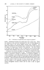
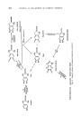
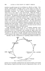
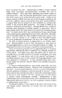
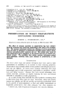
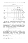
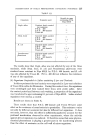
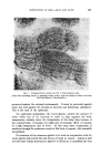
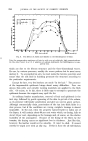
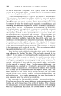
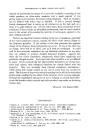
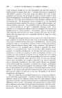
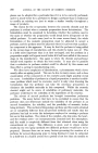
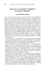
200 JOURNAL OF THE SOCIETY OF COSMETIC CHEMISTS permits these bottom-dwelling fishes to mimic accurately not only the color shade but also the pattern of their backgrounds. Experimentally, even a checkerboard pattern can be remarkably wall duplicated. • As a pigment, melanin also serves to offer some protection against the harmful effects of excessive sunlight radiation and is an essential component for efficient func- tion of the eye. In man, variations in melanin pigmentation have, in addition, acquired well-known cosmetic and social significance. In the full-blooded negro adult man the total amount of melanin by weight in the body has been estimated to be about one gram. • In all vertebrates, melanin formation is believed by most observers to take place about certain granules primarily in the cytoplasm of specializeinappear dendritic cells called melanocytes. In human skin, melanocytes routinely fixed and stained histologic sections as regularly spaced, individual, vacuolated or clear cells in or just adjacently below the basal layer of the epidermis and in the hair bulbs. These specialized cells arise embryologically from the neural crest. Several techniques can be used to visualize them clearly, with their long, slender, pseudopod-like processes or dendrites. They may be stained with silver, gold or methylene blue, or demonstrisofhisto-whichsolutionsintyrosinase, chemically after incubation under proper circumstances tyrosine or dihydroxyphenylalanine (dopa). 7 The enzyme : .... J• • _ •:: • ' r 4/1•,- ' ' -'•' '--' ' :• ß • ' ' • ' • ' - •' •- ' -" ':: :"• ' • , •.•.. . . . • ,• i• ' •' i•.•:.-- ...:•..,• . -: -• L• •---% •'.: •?-•: • . •' •:•,' ,,i*:!•.•':•..•. ' --,• ..... -'.•. ': •i• *..}:.s.•... •. • . '- '•'.-'... •,• - • •-:-- •,• •.• --: -•. • -.' ?:. :'• • -- '•:•q. --'.. .... - .-?'4• - .- •.' --.• -.•'--.- -,:-•-•-,' .. • '• :•. • •- -•,.: ...•:• - - -.•.• ." • ---.. •.,-? • - .: •. • .-:•. ,•:½ ,- . :•-• -- .•-- -- - •? ,?,s•- . - ... - "'•.•. -.- . -- ..• •-.• :/ - .• :: ........ -•,:•. -•...• .•,• :, t:-•F•.. -•• •:•' • ..- '.• -. •' '-'" i' ' * -•:• •'•[.' i:•:, "'•: ':: . .... '. ' '2 "•-•' --.':- • :. •.:•- :-• -•% • r • •d' •:" ::, • .:} • •.. -(.. ' . -' - .' : •..": •:.•' r• .-' cc • ...•. : • •- * • J' .. •-• . ..-•:• :• ..... -•.. •%•-•.. •. .:..:.•-•:• .•..'•.. .:-•.,-•..•, :'•.- .•..•:• ..• ...... g :-• -•:•. •,-:% .'-•:-i•.. •---•-. -.:"•- . .... •.•,• t• . .•?•. * • •5 .... . .• •'. . .: .:• • -. • . ....• . - . , •, • ..•.. •: • % -• •.•' • Fig. 2. Dermal surface of separated human epidermis prepared by dopa technique. ? present on special cytoplasmic granules in most melanocytes, catalyzes-the oxidation of tyrosine or dopa to the dark pigment melanin. In Figure 2 the dopa reaction has been used to demonstrate clearly these specialized cells in human skin. Apparently, there is more branching of the cells in a
Purchased for the exclusive use of nofirst nolast (unknown)
From: SCC Media Library & Resource Center (library.scconline.org)