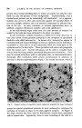
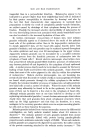
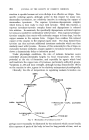
SKIN AND HAIR PIGMENTATION 201 tangential than in a perpendicular direction. Melanocytes appear to be hydrated to a greater degree than their neighboring basal cells as indicated by their greater susceptibility to destruction by freezing 8 and also by the fact that their characteristic clear appearance in routinely fixed preparations is chiefly the result of cytoplasmic pseudo-vacuole formation, an artifact caused by shrinkage of their cytoplasm during the process of fixation. Possibly the increased selective destructibility of melanocytes by the very short-lasting intense heat associated with atomic-bomb flash burns 9 can also be related to the increased hydration of these cells. In routine, microscopic cross-sections of human skin, most melanin granules ordinarily appear as if clustered above the nuclei of the palisade basal cells of the epidermis rather than in the dopa-positive melanocytes. In deeply pigmented skin, all the basal cells appear heavily laden with granules of melanin, and such granules may be scattered upward throughout the entire epidermis and may even fill macrophages in the upper corium. Apparently melanin granules can be inoculated by a so-called cytocrine secretory process from the dendritic processes of melanocytes into the cytoplasm of basal cells.•0 Recent electron microscopic observations show that actual bits of melanin-granule-filled dendritic processes of melanocytes may become pinched off and deposited in the cytoplasm of basal epidermal cells. A similar process clearly needs to be invoked to account for the heavy loading of hair shafts and feathers with melanin granules. Some observers claim that most of the active pigment in the skin lies within the cytoplasm of melanocytes. 7 Modern electron microscopists, too, are becoming less certain of just what lies inside or outside of cells as various systems of tubules are found which permeate through the cellular cytoplasm to the surface. Possibly the terminal dendritic processes of melanocytes actually penetrate into basal cells as such a system of tubules. Wherever most of the melanin granules may ultimately be found to lie in the epidermis, it is clear that some of them can be found in a free state in the cytoplasm of basal cells. Although melanin granules were at one time believed to develop as self- replicating mitochondrial particles, • recent electron microscopic observations clearly show that such granules form about cytoplasmic structures which make up the Golgi apparatus TM and that they are therefore, in a sense, analogous to the secretory granules which occur in enzyme-secreting glandu- lar cells such as those found in the pancreas. Melanin granules isolated from various mammalian sources by differential centrifugation are fairly uniform rounded bodies about 0.3 microns in diameter." The melanin polymer of such granules isolated from mouse melanomas is chemically linked with protein having pseudo-globulin properties and a minimum molecular weight of 20,000. When such melanin- protein granules are progressively digested by pancreatin, the sulfur content of the protein remaining attached to the melanin moiety increases. • This

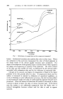
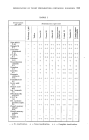
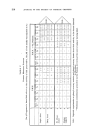
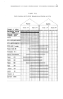
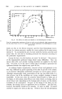
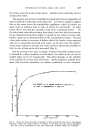
202 JOURNAL OF THE SOCIETY OF COSMETIC CHEMISTS ¸ ¸ / / / Relatively slow
Purchased for the exclusive use of nofirst nolast (unknown)
From: SCC Media Library & Resource Center (library.scconline.org)