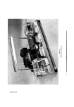
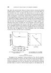
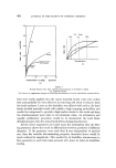
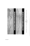
THE MELANOCYTE SYSTEM AND KERATINIZATION 415 epidermis appears as increasing shades of brown to black, whereas the same pigment deep in the dermis appears blue. The blue naevus and mon- golian spot are examples of this apparent blue colour of deeply situated melanin. The determination of natural skin colour The accurate matching, precise determination and recording of skin is difficult in dermatological practice. Photographic methods, using strictly controlled colour temperature lighting, with the emulsion developed under standardized conditions together with colour charts photographed alongside the subject, often fail to give satisfactory comparisons. A comparator method has also been used in which the skin is illuminated at the same time as a series of standard colours and the disc rotated until visual matching is obtained. Probably the best method is by the use of reflectance photometry the apparatus employed for this purpose is manufactured by Evans Electro- selenium Ltd. The principle of this technique depends on the absorption of rays of a selected wavelength directed on to the skin surface. The wave- length of the incident radiation is determined by interposing filters between the light source and the skin. The degree of absorption is estimated by measuring the light reflected back from the skin by means of a selenium cell. The current generated in the cell is recorded by a sensitive galvano- meter. The greater the quantity of the rays absorbed by the skin, the less radiation will be reflected back to the selenium cell and consequently the lower will be the galvanometer reading. For the determination of red due to blood in the skin, red light is suitable because in this region of the spectrum a good haemoglobin has a good maximum absorption. However, the maximum absorption by melanin occurs in the uv region of the spectrum and therefore the determination of the degree of brownness due to melanin was unsatisfactory when visible light was used with any of the filters supplied with the original apparatus. We therefore modified the photometer for use with uv rays. A uv light source in the form of an "overrun" projection lamp (Philips A1- 186) with a 1 mm Chance OX1 filter was substituted for the original light source to produce a radiation having a wavelength of around 13,600 21,. These rays are readily absorbed by melanin and because the selenium cell is sensitive to this range of uv light this gave a much more sensitive method of measuring the brown pigmentation of the skin (2).

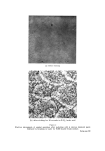
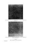
416 JOURNAL OF THE SOCIETY OF COSMETIC CHEMISTS A photographic technique was also developed in which a uniform radiation of the skin with uv rays was attained by a battery of four uv lamps (Osram 125 MEW/V) having a total output of about 14 watts at a wavelength of about 3,650 A. The area of skin under investigation was photographed through an OX1 1 mm filter at a standard distance and exposure, and the emulsion processed to a given gamma. Densitometry readings of the negative, compared with a simultaneously photographed control, were made with a radiological densitometer. This method gave comparable results with those obtained by reflectance photospectometry. THE NATURE OF SKIN PIGMENT Melanin, as already mentioned, is formed by specific epidermal cells known as melanocytes. The only criterion of a melanocyte is that it produces melanin, and at present the only satisfactory method for its histo- logical demonstration is by incubating epidermis in dihydroxyphenylalanine (dopa). It is remarkable that this compound should be more readily acted upon by the enzymes of the melanocyte than the postulated natural substrate, tyrosine, which is thought to be the true precursor of melanin. The reason given for this anomaly is that in vitro the melanocyte is unable to accomplish the first stage of transforming tyrosine into "dopa" and if, therefore, "dopa" is presented to the cell the rest of the metabolic pathway towards melanin formation proceeds to completion. As mentioned above, the precursor of melanin is thought to be tyro- sine, and by a series of reactions, some enzymatic oxidations by the enzyme tyrosinase and some autoreductions, this amino acid is transformed into the brown-black pigment, melanin. In the final product this is closely associated with protein to form melanin granules, which are then passed into adjacent epidermal cells. Another type of melanin has also been described, known as pheomelanin (3). This is the yellow-coloured pigment which is thought to be responsible for the yellow skin colour of the Oriental races and also for the yellow colour in the hair of "agouti" varieties of several animal species. Some authorities consider that the production of pheomelanin involves similar metabolic pathways to those of melanin synthesis and that possibly it is derived from the same precursor, tyrosine. A difference between these two melanins is shown by their solubilities in sodium hydroxide. It has also been suggested that pheomelanin represents a further oxidation product of melanin.
Purchased for the exclusive use of nofirst nolast (unknown)
From: SCC Media Library & Resource Center (library.scconline.org)