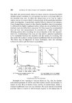
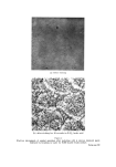
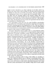
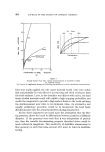
THE MELANOCYTE SYSTEM AND KERATINIZATION 417 OTHER ASPECTS OF MELANOCYTE FUNCTION The definition of a melanocyte is that it is a cell that forms melanin and is able to oxidize dihydroxyphenylalanine to a black pigment. It has also been shown that dendritic cells could be detected at higher levels in the epidermis than the basal layer. These cells situated in the upper regions of the epidermis were, however, dopa negative. Therefore other methods had to be used for their demonstration and the gold impregnation tech- niques were probably the most reliable for their detection. Because these so-called high-level melanocytes were dopa-negative it was considered that they were melanogenically effete and had no further function. They were carried passively upwards with the general movement of the epidermal cells, and were finally exfoliated with the keratin layer. Recently we have used various enzyme techniques on human and animal skins and were surprised to find that although these high level cells were inactive in respect to dihydroxyphenylalanine, nevertheless they were very active in respect to other substrates. The first enzyme to be detected was adenosine triphosphatase (5). Later we obtained evidence that they also exhibit acid phosphatase and sulphatase activity. It would, therefore, appear that while they no longer produce pigment they almost certainly perform other functions at higher levels in the epidermis. During the process of keratinization the epidermal cells, or keratino- cytes as they are sometimes called, become transformed into a stable, chemically-resistant fibrous protein known as keratin. This unreactive material, together with its contained lipids, acts as the first barrier against physical and chemical insults to the skin in particular, and to the body as a whole. The process of keratin formation appears to be a two-fold process. One is cellular cytolysis in which the nucleus and other components of the cell are removed before the occurrence of the second stage of molec- ular keratin polymerization. Thus, the physical nature of the final keratin layer can be modified to the particular site rcquirements of the animal depending on the relative amounts of the celhflar •naterial removed before keratinization (6). It is quite obvious that the horny layer on our palms and soles is differ- ent from that of other regions, but it can be shown that more subtle differences also exist between other skin sites. It is difficult to explain the mechanism whereby these regional differences in keratinization are brought

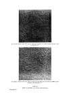
418 JOURNAL OF THE SOCIETY OF COSMETIC CHEMISTS about, but one possible means of affecting this would be by the action of these high-level dendrocytic cells. It is beyond reasonable doubt that the autolysis of the cell protoplasm prior to keratinization is due to hydrolytic enzymes released in the upper layers of the epidermis at the level of cells known as the granular layer. In skin diseases where there is no organised liberation of these enzymes, none of the cell contents is removed, and the whole of the cell, including its nucleus, becomes involved in the process of keratinization. Such a state of affairs exists in the skin disorder, psoriasis. Palmar and plantar keratinization occupy a position midway between the keratin produced in psoriasis and that, for example, produced on our backs. Here much of the cell contents, except the nucleus, escape auto- lysis and consequently the keratin layer is more solid and stable than that formed on the back. It is important to mention that although the keratin layer is different this does. not necessarily imply that the actual keratin molecules forming the horny layer differ from each other. As we have detected three distinct enzyme systems in melanocytes other than their dopa oxidase activity, it would perhaps seem reasonable that this other hydrolytic activity might influence the mode of keratiniza- tion of the epidermal cells. The ATPase activity is probably related to motility of the cells. It is most unlikely that it is the type of ATPase associated with ion transfer through cell membranes, and is probably the type associated with contrac- tile protein. This would suggest that the dendritic cells move actively up within the epidermis and are not passively carried upwards by the general movement of the epidermal cells. Acid phosphatase and sulphatase are both lysosomal types of hydro- lytic enzymes, and both would be capable of causing cellular hydrolysis if they were transferred into the epidermal cell in a similar manner to that in which the melanin granules are transferred into epidermal cells of the basal region. It is also possible that any effects of these high-level den- drites could be due to an induction or activation of hydrolytic enzymes rather than to an actual transfer of such an enzyme into epidermal cells. Although the precise mechanism cannot be directly demonstrated, the circumstantial evidence points strongly towards the involvement of the high-level dendrocytes. Thus in plantar skin where there is relatively little hydrolysis, it is extremely difficult to demonstrate ATPase active dendritic cells, whereas in normal epidermis where there is a much greater hydrolysis of the epidermal cells they are readily detectable in considerable numbers.
Purchased for the exclusive use of nofirst nolast (unknown)
From: SCC Media Library & Resource Center (library.scconline.org)