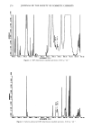
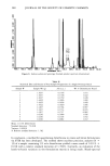
ALKANOL PERMEATION OF HAIRLESS MOUSE SKIN 239 was about 0.6 cm 2. After assembly, each chamber was filled with 1.4 ml of normal saline and placed in a constant temperature bath. The contents of both chambers of the cell were stirred at 150 rpm. It was shown that the permeability of this skin to certain alkanols increased gradually over the first ten hours of incubation in the diffusion cell (15). Therefore, skin sections in this study were equilibrated with saline for --12 hours to obtain a stable condition of hydration before beginning an experiment. The half-cell contents were then evacuated with a syringe, rinsed twice with normal saline, and filled with 1.4 ml of saline. After waiting for five minutes for the temperature to re-equilibrate, 200 txl of saline were placed in the receiver chamber (dermis side) and 100 }xl samples of stock solutions of two differently radio-labeled (3H and14C) permeants were placed in the donor com- partment (stratum corneum side). Although charging the donor half-cell began a run, the initial donor sample was not taken until --2 minutes had elapsed to allow for thorough mixing. Samples from the receiver chamber were withdrawn at 1000-second intervals for 7000 seconds, at which time a run was terminated. The overall experi- mental design and sampling schedule led to a pseudo-steady state for the permeation process. In order to get a complete homolog profile on each skin, runs were performed serially. Between runs the cell compartments were evacuated with a syringe equipped with a flexible polyethylene tubule, rinsed three times, and refilled with saline. The triple rinse was repeated approximately on the half hour twice more. A fourth single rinse about 30 minutes later completed the clean-up procedure. The permeability coefficients of all six solutes were determined on each skin by carrying out four sequential exper- iments over a total period of about 20 hours, excluding the initial 12-hour hydration. The order of running these experiments was: first run.' 3H-water and14C-ethanol second run.' 3H-methanol and•4C-butanol third run.' 3H-methanol and•4C-hexanol and fourth run.' 3H-methanol and•4C-octanol. The previously described dual-label procedure (15) showed that the permeabilities of the co-permeants are unaffected by each other at the solute concentrations used. It also offers obvious experimental efficiencies. Since the sequential runs included either methanol or water, two solutes with permeability coef- ficients highly sensitive to the stratum corneum's integrity, their permeation rates served as a check on a membrane's condition throughout an experiment. Some of the data used in the analysis for animals 60 to 120 days of age was excerpted from earlier studies not explicitly aimed at aging influences (15). These data are only included where no essential detail of the experimental procedures were at variance with the present protocol. DATA ANALYSIS Plots of receiver concentration (counts/volume) against time were made. The perme- ability coefficient is defined by (15): JT = P'A'AC - dM at (Equation 1) where JT = the pseudo steady state flux (cpm/hr), or the instantaneous amount pen- etrating the total membrane with time, dM/dt P = the permeability coefficient

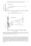
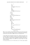
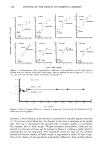
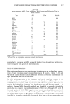
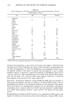
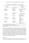
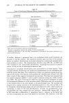
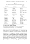
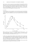
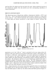
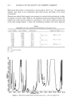
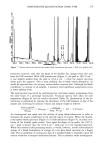
240 JOURNAL OF THE SOCIETY OF COSMETIC CHEMISTS (cm/hr) A = the diffusional area (cm2) and AC = the concentration differential across the membrane. This was taken to be equal to the donor phase concentration (cpm/ cm3). Since JT = VR ' (dC/dt), the permeability coefficient can be calculated from: p _ \ dt ,/ (Equation 2) A ß AC where V•t = the receiver half-cell volume (cm3), and (dC/dt) = the pseudo-steady state slope of the concentration versus time plot (cpm/cm3/hr). THICKNESS AND WEIGHT MEASUREMENTS Thickness of the skins was determined over the first year of mouse life. An excised dorsal or abdominal section several cm 2 in area was sandwiched between glass micro- scope slides. Using a micrometer, the thickness of the sandwich was measured with and without skin, the difference being the net thickness of the skin. The measurements were unaffected by varying pressure of the micrometer on the glass plates. Measurements were made in triplicate except at 108,328, and 342 days, where only two measurements were taken. No attempt was made to determine if soaking in the diffusion cells altered thickness. Since hydration effects on permeability are within the ultrathin stratum corneum for the most part, changes which might be noted would not have been relatable to permeability (to the stratum corneum). As a routine, mice were weighed before sacrifice. A number of weights were recorded at several ages and these exhibited little variability. For instance, six values were obtained at 25 days of age with an average weight of 14.2 gm and a standard deviation of 1.2 gm. RESULTS Several reports have appeared in the literature describing morphological changes that occur in the hairless mouse during the first weeks of life (16-20). The animals are born hairless but grow a coat of hair in the first few days after birth. This coat of hair appears normal up to the age of about 13 days, whereupon hair loss begins around the eyes and the nose, and then, in caudal progression, all hair is shed. At 21 days of age the body is virtually nude and covered with a thin, smooth, and pinkish skin. Around 35 days, a sparse second growth of hair often appears, which is lost between 45-60 days of age. The skin retains its "young appearance" for several months, but it gradually assumes a greyish cast and takes a grainy texture. Past 6 months of age the animals show obvious signs of aging. The skin develops wrinkles, in some cases forming deep folds, and its color darkens. The animals concomitantly become less active. Figure ! depicts mice weights as a function of age. A total of 224 weight measurements are summarized in this graph. Skin thicknesses of mice ranging in age from 2 days to 342 days are graphically presented in Figure 2. Average thicknesses have been plotted along with standard deviations at each measurement point. Permeability coefficients of water, methanol, ethanol, n-butanol, n-hexanol, and n- octanol determined on abdominal and dorsal skins of mice 4 days to 360 days of age
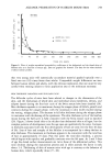
Purchased for the exclusive use of nofirst nolast (unknown)
From: SCC Media Library & Resource Center (library.scconline.org)