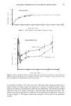
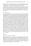
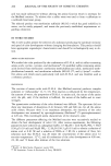
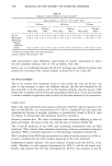
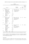
274 JOURNAL OF THE SOCIETY OF COSMETIC CHEMISTS USP reference standard polydimethysiloxane (F-3, U.S.P.C., Inc.) to a final volume of 50.00 ml with methylene chloride (#9315-3, J. T. Baker Chemical Company). A 0.001-g/ml dimethicone working standard was then prepared by volumetrically di- luting a 1.00-ml aliquot of the stock solution to 10.00 ml with methylene chloride. A 40% v/v hydrochloric acid solution needed for the solvent extraction step was pre- pared from concentrated hydrochloric acid (#2062, Mallinckrodt, Inc.) and distilled water (Purified Water, U.S.P., Electrified Water Company). APPARATUS Instrumental analysis was performed on a Nicolet 5MX Fourier transform infrared spectrometer with 4-cm-1 resolution (Nicolet Instrument Corp.) attached to an X-Y recorder (Model 7010B, Hewlett-Packard) and interfaced to a diskette storage drive (Model SA 800/801, Shugart). The sample cell used was a demountable liquid cell with 0.5 mm-Teflon spacers (Harrick Scientific Corp.) and sodium chloride transmis- sion windows (13 x 2mm, #7000-301, Barnes Analytical Division, Spectra-Tech, Inc.). The sample cell was filled using a glass syringe (#2140, Becton-Dickinson). SAMPLE PREPARATION 1. Extraction. Accurately weigh ca. 1.5 g of sample into a 50-ml graduated cylinder with ground glass stopper. Add about 20 ml of 40% v/v HC1 solution and pipette in 15.00 ml of methylene chloride. Shake vigorously, manually, for one minute and allow to separate into layers. 2. Separation. Pipette a 10.00-ml aliquot of the bottom, organic phase into a 10-ml disposable syringe (#5604, Becton-Dickinson) which is attached to an Acrodisc filter (0.45 •tm pore size, #4404, Gelman Sciences) which is, in turn, attached to a Silica SEP-PAK cartridge (#51900, Waters Associates, Inc.). Pass the aliquot and a 1.0-ml methylene chloride wash through the system, collecting the effluent in a 25-ml Erlen- meyer flask. 3. Final Solution. Reduce the volume of the collected fraction almost to dryness on a warm hot plate. Care should be taken to assure that the solution does not froth over. Take up the residue in methylene chloride and dilute, volumetrically, to 10.00 ml. ANALYSIS The empty liquid cell was scanned into the background file. (When plotted, all files are automatically ratioed against the background file.) The solvent, standard, and sample solutions were scanned into the remaining files. Ten scans were taken in each case, signal-averaged, and the absorbance spectra plotted on the X-Y recorder. In addition to plotting the full spectra, absorbance values at the peaks of interest were read directly from the digital display on the 5MX. A particularly useful feature of digital data handling in FTIR is the capability of performing spectral subtractions for example, the solvent portion may be eliminated from a solution spectrum. A difference factor, indicating the fraction of solvent in the solution, is entered, and the microprocessor then subtracts that proportion of the solvent

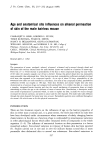
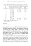
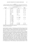
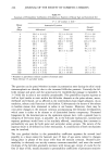
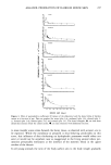
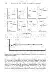
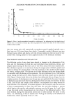
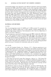
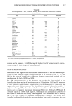
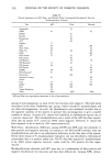
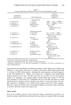
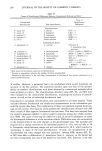
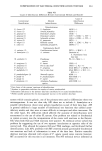
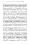
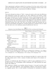
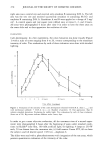
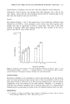
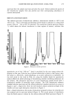
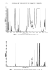
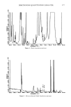
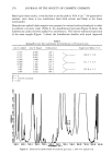
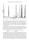
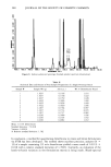
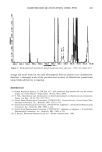
DIMETHICONE QUANTITATION USING FTIR 275 spectrum from the sample spectrum and plots the result. Solvent-subtracted spectra of the dimethicone samples were also plotted and their corresponding absorbances of interest determined. RESULTS AND DISCUSSION The infrared spectrum of dimethicone exhibits a characteristic doublet at 1097.6 and 1014.6 cm-1 due to stretching and bending of Si-O bonds in addition to a sharp band around 1260 cm-1 due to CH3-Si vibrations. The methylene chloride spectrum (Figure 1), which shows the solvent interference in these regions of interest, indicates that 4000.0 401•.0 •4OO.O •11•O.O Z•00.O 1BD0.O 1GIX].O l•OO.O ll]X].O OOO.OO 7120.O0 Figure 1. Methylene chloride spectrum. quantitative use of the 1260-cm-• band is precluded by the very strong solvent ab- sorption in that area. Even the use of solvent-subtraction is futile in that region since the solvent is, effectively, totally absorbing, an accurate subtraction is impossible. As was well noted in the work by Rihs (2), using the doublet bands for quantitative analysis affords the added advantage of determining whether interfering substances are also present. The absorption spectrum of a pure dimethicone sample should show a symmetrical doublet of essentially equal intensity. The USP dimethicone reference standard solution spectrum and the solvent-subtracted standard spectrum are given in Figures 2 and 3, respectively. Note the well resolved, almost symmetrical shape of the doublet and near equal absorption intensities in Figure 3. Next, a blank formulation (the product formulation prepared without the dimethicone) was analyzed and its spectrum plotted (Figure 4). The actual amount of interference from the blank matrix is shown by the solvent-subtracted blank spectrum (Figure 5).
Purchased for the exclusive use of nofirst nolast (unknown)
From: SCC Media Library & Resource Center (library.scconline.org)