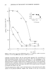
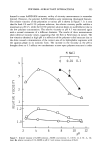
J. Soc. Cosmet. Chem., 41, 227-233 (July/August 1990) The simultaneous penetration of water and sodium lauryl sulfate through isolated human skin MARIE LOD•N, Medical Department, Research and Development, ACO AB, S-171 26 Solna, Sweden. Received September 28, 1989. Synopsis Surfactants can influence the barrier function of the skin and induce an increased transepidermal water loss. The simultaneous penetration of water and sodium lauryl sulfate (SLS) (0.1, 1.0, and 10.0% w/w) through isolated human skin was measured using tritiated water and 35S-labeled SLS. The amount of SLS penetrating the skin was 50-100 times higher from 1.0% SLS than from 0.1%. Increasing the concentration from 1.0 to 10% increased the amount penetrating ten times. The rate of penetration also increased with respect to time, indicating increasing damage to the skin over the duration of contact. Since no true steady state was obtained, only pseudopermeability constants, p-Kp, could be calculated (obtained by division of the penetration rate by the applied concentration). The p-Kp of SLS at the arbitrary times 21-24 hr was 0.1, 1.0, and 1.3 Ixcrn/min -• for 0.1, 1.0, and 10%, respectively. The p-Kp of water at the same arbitrary time was 55 •xcrn/min -• when 0.1% SLS was present and 120 Ixcm/min -• when 1.0 and 10% were present. The results indicate that skin damage from washing solutions is not linearily related to the surfactant concentration in the solution. The monomer activity of SLS was not constant above critical micelle concen- tration (CMC) but increases when the concentration of micelies increases. INTRODUCTION Human skin is daily exposed to surfactants. As a result of these exposures, the barrier function of the skin can be changed (1-6). For a certain surfactant, the degree of damage is dependent on the concentration in the skin, i.e., on the penetration rate. Sodium lauryl sulfate, SLS, is an anionic detergent frequently used in experimental dermatology to study the biological response to surfactants. Exposure of the skin to SLS changes the lipid composition of the stratum corneum (7). The composition of the intercellular lipids seems to be an important factor in the regulation of epidermal per- meability (8,9). SLS extracts lipids from the intercellular spaces in the stature corneum (10) and stimulates keratinization (11) and epidermal sterol and fatty acid biosynthesis (12). There are few data in the literature concerning the absolute amount of surfactant that will enter the skin upon exposure. It is believed that the present study is the first to 227

228 JOURNAL OF THE SOCIETY OF COSMETIC CHEMISTS quantify the penetration of SLS from aqueous solutions and correlate that to simulta- neous diffusion of water. MATERIALS AND METHODS CHEMICALS Sodium lauryl 35S-sulfate (35S-SLS) and 3H-water were obtained from Amersham, UK. 35S-SLS was purified prior to use to a radiochemical purity 97%, according to a method of Perrin et al. (13). The purity was determined by thin-layer chromatography on silica plates. The plates were developed in isopropanol:chloroform:methanol:water (40:40:20:8 v/v) or in chloroform:ethyl acetate:formic acid (50:40:10). The radioac- tivity on the plates was located using a Berthold LB 2723 radioscanner. Unlabeled SLS was obtained from Henkel Kemi, purity 98%. The donor solution consisted of 35S-SLS and unlabeled SLS dissolved in 3H-water. The SLS concentrations were 0.1, 1.0, and 10% (w/w), with a radiochemical concentration of 1 X 10 •ø Bq/ml. The radiochemical concentration of water was 7 X 10 lø Bq/ml. ANALYSES The radioactivity was measured in a Packard Tri-Carb liquid scintillation counter, model 3375, with Instagel (Packard) as scintillation cocktail. 35S and 3H have different energy levels, thus permitting simultaneous quantitation of SLS and water in the samples by measuring the radioactivity at different energy regions and correcting for quenching and spillover. The radiochemical concentration of the isotopes was chosen so that the number of counts of 35S in the lower energy region was low, compared with the counts due to tritium. PERMEABILITY EXPERIMENTS The penetration experiments were performed according to similar procedures described previously (14, 15,16). Human skin was obtained from cosmetic surgery at the Depart- ment of Plastic Surgery, Sabbatsbergs Hospital, Stockholm, Sweden. Normal skin from three females was used, one abdominal and two breast skin. Due to differences in size, the abdominal skin and one of the breast skin samples were divided into six smaller pieces each, whereas breast skin from the other donor was divided into three pieces. Five experiments were performed on each concentration, and the same combination of skin samples was used (i.e., two abdominal + two breast + one breast skin). After mechanical removal of the subcutaneous fat, pieces of skin were wrapped in aluminium foil and stored at -20øC. Harrison et al. reported that freezing of the skin did not affect the permeability (17). Before starting an experiment, samples were thawed at room temperature. Pieces of skin were mounted in flow-through diffusion cells of stainless steel. The diffu- sion cells were kept in a water bath at 30øC. The SLS solution, 200 Ixl, was applied to the outer surface of the skin. The area available for diffusion was 0.5 cm 2. Beneath the skin, through the lower section of the cell, a receptor medium (normal saline) was pumped and collected in polyethylene vials. The continuous supply (approximately 2
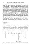
Purchased for the exclusive use of nofirst nolast (unknown)
From: SCC Media Library & Resource Center (library.scconline.org)