SKIN AND IMMUNITY 2 5 5 data confirm that LC are immune macrophages that provide an immune barrier in the epidermis. While LC have the capacity to produce IL-1, it has also been observed that keratino- cytes have the capacity to produce rather large amounts of a factor that was biologically indistinguishable from IL-1. This factor, termed epidermal cell-derived thymocyte-ac- rivating factor (ETAF), has biologic activities that mimic that of IL-1, indicating that the keratinocyte, believed to be a "non-immune" cell, has the capacity to elaborate at least one immunoregulatory factor, classifying it as an "immunoregulatory cell" (3,6). Not only do keratinocytes produce ETAF, an initiation of T-cell activation, but it has also been proposed that UVR-induced alteration in ETAF production may be respon- sible for several important biological effects of UVR (5). ETAF may even regulate the excessive proliferation of keratinocytes that occurs in psoriasis (14). There is decline of ETAF with age. ETAF represents another mechanism for immune compromise in old skin. The similarity of ETAF to IL-1 is so marked that they are often referred to as ETAF/IL-1 (15). Increase in ETAF occurs when stimulated by an irritant or antigen. Thus, while processing and presenting of an antigen is done principally by LC, they are possibly assisted by the keratinocyte that can elaborate ETAF/IL-1. The neuropeptide alpha-melanocyte-stimulating hormone (ot-MSH) can act as an an- tagonist to IL-1 bioactivities such as the inhibition of thymocyte proliferation. Recently it was reported that topical application of ot-MSH suppresses the cutaneous immune response to contact sensitizers. Further, the loss of contact hypersensitivity (CH) due to applications of ot-MSH could be reconstituted by intradermal or intravenous injections of ETAF/IL-1. Topical application of ot-MSH did not cause alterations of LC. These observations suggest that ot-MSH may represent an additional biologic modifier that can modulate suppression of the CH responses to various haptens (15). This model of epidermal function is made more complex by the observation that factors other than ETAF are also produced by keratinocytes. For example, interleukin-3 (IL-3) is a growth factor that is produced by activated T-lymphocytes and T-cell lines. Both human epidermal cells and squamous cell carcinoma cells have the capacity to produce IL-3. Moreover, there has been derived from epidermal cells a novel factor that inhibits in vitro hypoproliferative responses and IL-2 products (16). This factor, termed epi- dermal cell-derived lymphocyte-differentiating factor (ELDIF) is produced by keratino- cytes. Detailed discussion of the role of interleukins and interferons in immunological skin function is beyond the scope of this review. However, mention should be made that gamma interferon is a lymphokine secreted mainly by activated T-lymphocytes and capable of inducing a wide range of effects on many cells. IL-1 is secreted by macro- phages following uptake of antigen-antibody complexes or following contact with T- lymphocytes during antigen presentation. Secretion of IL-1 is enhanced by gamma in- terferon. IL-1 induces secretion of IL-2, which promotes the growth of T-cells after antigen presentation. IL-6 is a cytokine secreted by phagocytes and activated T-lym- phocytes, as well as by some nonlymphoid cells (e.g., fibroblasts). IL-6 was originally described as a factor that promotes the differentiation of B-lymphocytes to antibody secretory cells.

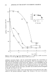
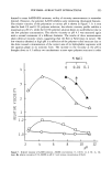
256 JOURNAL OF THE SOCIETY OF COSMETIC CHEMISTS ANTIOXIDANTS, AGING SKIN, UV, FATTY ACIDS Antioxidants (e.g., BHA, BHT) can play a role in altering immune responses. They can alter CH responses and, when applied topically to experimental animals, alter the den- sity of epidermal immune macrophages such as LC and T-cells. The alterations may be metabolized via the arachidonic acid (AA)-prostaglandin pathway. BHA has immuno- suppressant activity at levels that are not cytotoxic, affecting both B- and T-cell func- tion. In addition, LCs are subject to free radical attack, and their numbers are decreased via lipid peroxidation, probably through mediation via AA or its metabolites. Quenching of the free radical oxygen molecules in this pathway by antioxidants irre- versibly inhibits the cyclooxygenase enzyme, thereby affecting AA metabolism directly and/or indirectly via oxidation of AA into its products, prostaglandin, and leukotrienes (9,17). In aging skin, not only is there a decrease in ETAF and T-cells, but also in LC respon- sible for recognition of foreign antigens. There is an age-associated decrease in delayed hypersensitivity in human skin. The decrease reflects, in part, the decrease in total number of circulating thymus-derived lymphocytes (T-cells) and in their responsiveness to standard mitogens (mitotic-producing agents). In addition, epidermal LC, the cell population believed to be responsible for recognition of foreign antigens, have been reported to decline during adulthood in sun-exposed sites, with further loss in habitu- ally sun-exposed skin of older people. This exaggeration of age-associated cell loss in habitually sun-exposed skin may be relevant to photocarcinogenesis in the elderly. The loss of LC would be expected to impair the skin's cell-mediated immune response, particularly contact sensitization, because these cells are necessary for processing foreign antigens. It seems likely that there is the induction of some biochemical transducer in the skin that converts the physical stimulus, i.e., ultraviolet light, into a biochemical signal (18). Studies have documented the presence of prostaglandin synthetase and active synthesis of prostaglandins in the skin of various animal species. Various stimuli, such as me- chanical trauma and physical injury, including UVB irradiation, activate phospholipase A2 and the oxidation of arachidonic acid (AA) to prostaglandins. There is increasing evidence to implicate prostaglandins (PGs), leukotrienes (LTs), and other related lipids in the regulation of immune cells. AA has a biphasic effect on the number and functions of LC. Low topical doses of AA increased LCs. Higher doses (2%) decreased LCs and suppressed skin immune reactions in animal models. The increase may be correlated with increased immune reactivity and the decrease with a suppressed immune response (19). Oleic acid and linoleic acid (LA) were tested. Linoleic acid, but not oleic acid, can be converted by cells into arachidonic acid. Oleic acid caused a modest depletion of LC. Linoleic acid caused a much greater depletion, comparable to the effects observed with AA. All three acids caused hyperplasia (increase in number of cells) of the keratinocytes. AA and LA caused an increase in the number of mice epidermal pigment cells, and both induced melanogenesis (20). As previously noted, UV light and AA alter the cutaneous immune response, possibly by production of prostaglandins (PG). Of the PGs, only PGE2 suppressed the response of the skin to contact allergens. This suggests a specific role for PGE2 in the skin immune response. It is also possible that PGE2 affects epi-
Purchased for the exclusive use of nofirst nolast (unknown)
From: SCC Media Library & Resource Center (library.scconline.org)