IN VIVO PHOTOPROTECTIVE EFFECT OF CEG 309 output peak at 302 nm. The flux rate measured at the skin surface was 0.80 mW/cm 2. For each subject, the minimal erythema dose (MED) was determined preliminarily, and an irradiation dose corresponding to the double of the MED was used throughout the study. For each subject, eight sites on the ventral surface of one forearm and six sites on the other were defined using a circular template (1 cm 2) and demarcated with permanent ink. Two different formulations of CEG were used: a CEG-saturated solution in dis- tilled water and a CEG-saturated solution in water:Arlasolve DMI 50:50. A solution consisting of water:Arlasolve DMI 50:50 (without CEG) was also used to assess the DMI effect on UVB-induced skin erythema. For each subject, two skin sites were left untreated but exposed to UVB radiation (control). The protocol consisted of two series of experiments (6). In the first series (pretreatment protocol), the formulations tested were applied randomly for 3 h on the skin sites of one forearm using a Hill Top chamber (Hill Top Research Inc., Cincinnati, OH) whose cot- ton pad was saturated with 150 ptl of the formulation being tested. After 3 h, the chambers were removed, the skin surfaces were gently washed with water to remove the formulation, and each pretreated site was exposed to UVB irradiation. The second series of experiments was simultaneously performed on the other forearm of the same subjects. Skin sites were exposed to UVB irradiation, and then the formula- tions tested (150 ptl) were immediately applied to the irradiated sites (using the same Hill Top chamber described above) for 6 h. After this period, the chambers and the for- mulations were removed. For both experimental protocols, UVB-induced erythema was monitored for 58 h using the reflectance spectrophotometer described above. From the skin spectral data obtained, the erythema index (E.I.) was calculated using equation 1 reported by Dawson et al. (7): 1 1 1 1 1 E.I. --- 100 [Log -- + 1.5 (Log-- + Log ) -2 (Log-- + Log )] (1) R560 R540 R580 Rs: 0 R610 where 1/R is the inverse reflectance at a specific wavelength (560, 540, 580, 510, and 610 nm). E.I. baseline values were taken at each designated site before application of the formulations tested (pretreatment protocol) or before UVB irradiation (posttreat- ment protocol), and they were subtracted from the E.I. values obtained after formula- tion application at each time point, to determine A E.I. values. For each site, the area under the response (AE.I.)-time curve (AUC) was computed using the trapezoidal rule. AUC values were inversely related to the ability of the formulations tested to inhibit UVB skin erythema. To better compare the efficacy of the different formulations tested, the percentage inhibition of UVB skin erythema (PIE) was calculated from AUC values using the following equation: AUC(c ) - AUC(T ) Inhibition % (PIE) - X 100 (2) AUC(c ) where AUC(c ) is the area under the response-time curve of sites that received no treat- ment (control), AUC(T ) is the area under the response-time curve of the sites treated with the formulations being tested. Statistical analysis of the results were performed us- ing Student's t-test.

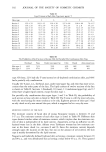
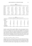
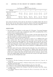
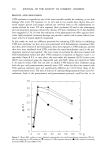
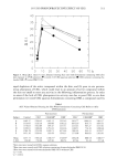
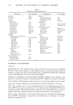
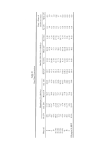
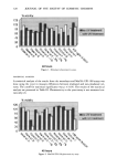
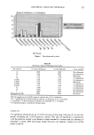
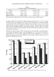
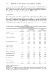
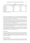
310 JOURNAL OF THE SOCIETY OF COSMETIC CHEMISTS RESULTS AND DISCUSSION UVB erythema is regarded as one of the most suitable models for studying in viva skin damage after acute UV exposure (1). In vitro and in viva studies have shown that acti- vated oxygen species and oxygen radicals are involved both in the inflammatory re- sponse elicited by acute UV skin exposure (skin erythema) (8) and in the photoaging and carcinogenesis processes induced by chronic UV skin irradiation (9). It has therefore been suggested (1,10,11) that the evaluation of the photoprotective effect against ultra- violet light-mediated cutaneous damage can provide a useful tool to assess radical scav- enger activity of topical applied compounds. In this study, we used two different protocols for evaluating CEG ability in inhibiting UVB skin erythema: (a) skin sites were pretreated with formulations containing CEG, and then, after removal of the formulation, they were exposed to UVB radiation, and (b) skin sites were irradiated with UVB, and then the same formulations used in the pre- treatment protocol were applied. The time course of erythema for skin sites treated with CEG formulations before and after UVB irradiation is reported in Figures 2 and 3, re- spectively. From A E.I. vs time plots, the area under the response (A E.I.)-time curve (AUC) was computed using the trapezoidal rule, and AUC values are reported in Table I. As may be noted, CEG was not able to inhibit UVB-induced skin erythema using both the pre- and posttreatment protocols since AUC values for skin sites treated with CEG aqueous solutions were not significantly different (p 0.05) from those of the control (untreated sites). The lack of activity of carboxyethylgermanium from water for- mulations, both in the pretreatment and posttreatment protocol, could be due to: (a) 55 50 25 . 20 :1 q$ 0 10 20 50 40 50 60 70 h Figure 2. Mean AE.I. values vs time, obtained treating skin sites with formulations containing CEG fore skin exposure to UVB radiation: (•') control (O) CEG aqueous solution (O) CEG solution containing Ariasolve DMI (V)water:DMI 50:50.
Purchased for the exclusive use of nofirst nolast (unknown)
From: SCC Media Library & Resource Center (library.scconline.org)