IN VIVO PHOTOPROTECTIVE EFFECT OF CEG 313 plied before skin exposure to radiation. We conclude that topical application of CEG formulations containing DMI could be used for skin care products containing radical scavengers. Further in vivo studies are planned in order to compare radical scavenger activity of CEG with that of others generally used in cosmetics and to assess the effect of different skin penetration enhancers on CEG photoprotection. REFERENCES (1) D. L. Bissett, R. Chaterjee, and D. P. Harmon, Photoprotective effect of superoxide-scavenging anti- oxidants against ultraviolet radiation-induced skin damage in the hairless mouse, Photodermatol. Pho- toimmunol. Photoreed, 7, 56-62 (1990). (2) J. R. Trevithick, Topical composition containing tocopherol esters of p-carotene for treating sunburns, PCT Int. AppL WO 93 03,720 1-27. (3) C. Duval, P. Lange, and M. C. Podman, Inhibition of cutaneous inflammation by free radical scav- engers. Proceedings of the 17th IFSCC Congress, Yokohama, 1992, pp. 453-461. (4) C. Guyomard, C. Chesne, I. Morel, and J. Cillard, Activite antiradicalaire du sesquioxyde de germa- nium, Parrums, Cosmdtiques, Aromes, 113, 45-46 (1993). (5) Test report by BIOPREDIC 1993: Protection de cellules diff•renci•es par des d•riv•s du germanlure vis-a-vis des m•tabolites toxiques de l'oxyg•ne (hydroperoxyde formation and lipid diene formation in rat hepatocyte cell cultures). (6) L. Montenegro, E Bonina, L. Rigano, S. Giogilli, and S. Sirigu, Protective effect evaluation of free rad- ical scavengers on UVB induced human cutaneous erythema by skin reflectance spectrophotometry, Int. J. Cosmet. Sci., 17, 91-103 (1995). (7) J. B. Dawson, D. J. Barker, D. J. Ellis, E. Grassam, J. A. Carterill, G. W. Fisher, and J. W. Feather. A theoretical and experimental study of light absorption and scattering by "in vivo" skin, Phys. Med. BioL, 25,696-709 (1980). (8) H. S. Black, Potential involvement of free radical reaction in ultraviolet light-mediated cutaneous damage, Photothem. PhotobioL, 46, 213-221 (1987). (9) E Urbach and P. D. Forbes, "Photocarcinogenesis," in Dermatology in General Medicine, T. B. Fitz- patrick, A. Z. Eisen, K. Wolff, I. M. Freedberg, and K. E Austen, Eds. (McGraw Hill, New York, 1987), pp. 1475-1480. (10) J. P. Cesarini, P. Miska, and M. C. Podman, The effect of antioxidants on human's erythema and sun- burn cells., Photothem. PhotobioL, 47, 73S (1988). (11) D. Dart, S. Coombs, S. Dunsrant, T. Manning, and S. Pinnell, Topical vitamin C treatment inhibits ultraviolet radiation-induced damage to porcine skin,J. Invest. Dermatol., 96, 353 (1991). (12) S. Kumar, A. W. Malick, N.M. Meltzer, J. D. Mouskountakis, and C. R. Behl, Studies of in vitro skin permeation and retention of a leukotriene antagonist from topical vehicles with hairless guinea pig model, J. Pharm. Sci., 81,631-634 (1992). (13) C. R. Behl, H. Char, S. B. Patel, D. B. Mehta, D. Piemontese, and W. Malick, "In Vivo and In Vitro Uptake and Permeation Studies," in Topical Drug Bioavailability, Bioequivalence, and Penetration, V. P. Shah and H. I. Maibach, Eds. (Plenum Press, New York, 1993), pp. 225-259.

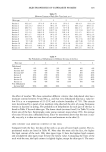
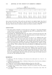
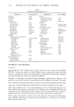
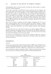
j. Soc. Cosmet. Chem., 47, 315-323 (September/October 1996) A comparison of two new in vitro phototoxicity methods to a published yeast phototoxicity method S. R. RACHUI, T. BOUFAISSAL, E. A. NEWCOMBE, and R. J. ALLEN, Stephens 6- Associates, Inc., 3310 Keller Springs Road, Suite 130, Carrollton, TX 75006 (S.R.R., T.B., E.A.N), and Shaklee Corporation, 1992 Alpine Way, Hayward, CA 94545 (R.J.A). Accepted for publication September 30, 1996. Synopsis Two new in vitro phototoxicity methods were compared to a previously published method that utilized yeast as a phototoxicity indicator. This comparison was done with eleven materials, tested in each system, to determine assay correlation. The protocols for the first two systems were similar and measured statistical differences in cell viability between irradiated and non-irradiated cell culture as a phototoxicity endpoint. The endpoint in the yeast model was a zone of inhibition generated in response to dosed phototoxic materials. The experimental sys- tems chosen for this work were (a) a monolayer of human neonatal fibroblasts and (b) the MatTek EPI-100 test system. UVA was used in these methods to elicit phototoxic responses from the test materials. It appears the MatTek EPI-100 system may be a more realistic predictor of phototoxicity as replicate standard deviations are smaller and statistically significant differences are more easily obtained. The MatTek system also has the advan- tage of being more similar to human skin than the other tested models. The monolayer system, however, may be the most sensitive of the models due to test materials directly contacting the cells and not being restricted by a stratum corneum, such as with the MatTek system. However, since statistical significance is more difficult to achieve due to greater inherent variability. The monolayer system may better be used as a screening tool. The yeast method may also be most useful when used as a rapid and inexpensive means of screening large numbers of materials prior to proceeding to more sophisticated and costly tests. INTRODUCTION Phototoxic materials are those that are not toxic under normal circumstances but that are chemically altered and become toxic when exposed to UV light. Evaluating phototoxicity, therefore, is an important addition to the toxicology profile of topically applied materials. A listing of materials that may stimulate phototoxic reactions is shown in Table I. In addi- tion, many materials that are known photoallergens may also be phototoxins. These materi- als include topical antimicrobials, fragrances, and sunscreen ingredients. Phototoxicity is normally evaluated in human subjects. This, however, can be costly and time-consuming when comparing multiple formulations. Using in vitro phototoxicity methods it is possible to evaluate materials quickly and accurately by measuring the product's effect on toxicity both before and after irradiation with UVA. UVA was se- lected as the irradiation source because most phototoxic materials respond to UVA (1). Research using a combination of UVA/UVB, such as with a solar simulator, was not done due to the strong cytotoxicity caused by UVB. This research compares two new as- says for phototoxicity evaluation to a previously published yeast method (2). 315
Purchased for the exclusive use of nofirst nolast (unknown)
From: SCC Media Library & Resource Center (library.scconline.org)