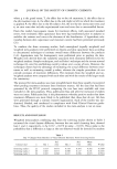
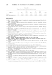
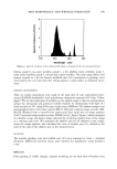
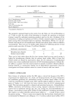
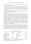
288 JOURNAL OF THE SOCIETY OF COSMETIC CHEMISTS Table III Changes From Baseline from the Definitive Study Std. Std. Mean Endpoint n Bar 1 error Bar 2 error delta P-value Dryness 99 1.041 0.028 1.235 0.028 0.194 .0001 Log10 (skin capacitance) 99 -0.023 0.003 -0.046 0.003 -0.023 .0001 REFERENCES (1) W. G. Cochran, Problems arising in the analysis of a series of similar experiments,•J. Roy. Stat. Soc. Suppl., 4, 102-118 (1937). (2) W. G. Cochran and G. M. Cox, Experimental Designs (John Wiley & Sons, Inc., New York, 1957), 115-129, 545-568. (3) R. B. D'Agostino and M. Weintraub, Meta-analysis: A method for synthesizing research, Clin. Phar- macol. Therapeut., 58, 606-616 (1995). (4) L. V. Hedges and I. Olkin, Statistical Analysis for Meta-Analysis (Academic Press, Orlando, 1985), pp. 27-117. (5) K. A. L'Abbe, A. S. Dersky, and K. O'Rourke, Meta-analysis in clinical research, Ann. Intern. Med., 107, 224-233 (1987). (6) T. A. Louis, H. V. Fineberg, and F. Mosteller, Findings for public health from meta-analyses, Ann. Rev. Publ. Health, 6, 1-20 (1985). (7) H. S. Sacks, J. Berrier, D. Reitman, V. A. Ancona-Berk, and T. C. Chalmers, Meta-analysis of ran- domized control trials, N. Engl. •J. Med., 316, 450-455 (1987). (8) S. B. Thacker, Meta-analysis: A quantitative approach to research integration,J. Am. Med. Assoc., 259, 1685-1689 (1988). (9) K. W. Wachter, Disturbed by meta-analysis?, Science, 241, 1407-1408 (1988). (10) N.M. Laird and F. Mosteller, Some statistical methods for combining experimental results, Intl. Technol. Assess. Health Care, 6, 5-30 (1990). (11) G. V. Glass, Primary, secondary, and meta-analysis of a large collection of research, Educ. Res., 5, 3-8 (1976). (12) I. Olkin, Keynote address: Meta-analysis: reconciling the results of independent studies, Stat. Med., 14, 457-472 (1995). (13) T. C. Chalmers, Problems induced by meta-analyses, Stat. Med., 10, 971-980 (1991). (14) J. C. Bailar III and F. Mosteller, Guidelines for statistical reporting in articles for medical journals: Amplifications and explanations, Ann, Intern. Med., 108, 266-273 (1988). (15) R.J. Light, Accumulating evidence from independent studies: What we can win and what we can lose, Stat. Med., 6, 221-228 (1987). (16) C. Mann, Meta-analysis in the breech, Science, 249, 476-480 (1990). (17) K.D. Ertel, B. H. Keswick, and P. B. Bryant, A forearm controlled application technique for esti- mating the relative mildness of personal cleansing products,.]. Soc. Cosmet. Chem., 46, 67-76 (1995). (18) B.J. Winer, Statistical Principles in Experimental Design (McGraw-Hill, New York, 1971), pp. 685-711, 397-398. (19) G. D. Steel and J. H. Torrie, Principals and Procedures of Statistics (McGraw-Hill, New York, 1960), pp. 146-159. (20) V. Rogiers, M.P. Derde, G. Verleye, and D. Roseeuw, Standardized conditions needed for skin surface hydradon measurements, Cosmet. Toiletr., 105, 73-82 (1990). (21) S. Dikstein, M. Katz, A. Zlotogorski, ¾. Broun, D. Wilson, and H. Maibach, Comparison of different instruments for measuring stratum corneum moisture content, Intl..]. Cosmet. Sci., 8, 289-292 (1986).

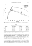
j. Soc. Cosmet. Chem., 48, 289-295 (November/December 1997) In vitro assay of high-SPF sunscreens R. P. STOKES and B. L. DIFFEY, Regional Medical Physics Department, Dryburn Hospital, Durham DH1 5TW, UK. Accepted for publication December 1, 1997. Synopsis In vitro spectral transmission measurements using excised human epidermis as the substrate were used to determine the photoprotection provided by physical and organic chemical sunscreens encompassing a wide range of sun protection factor (SPF). The measured SPFs were in good agreement with the quoted SPFs of the products. This in vitro technique using human epidermis could prove reliable for evaluating the SPF of high-protection sunscreens for which in vivo assay is problematic due to the impractically long irradiation times to achieve erythema on sunscreen-protected skin. We also compared our calculated SPFs, which assumed a natural sunlight spectrum, with those that would have been obtained assuming a xenon-arc solar simulator spectrum. We found that for products with relatively low UV-A absorption, the use of a solar ß simulator for in vivo testing overestimates the SPF that would be expected in sunlight. INTRODUCTION The photoprotection provided by a sunscreen product is assessed in terms of its sun protection factor (SPF). Sunscreen SPFs are generally measured by in vivo assay and defined as the ratio of the ultraviolet (UV) dose required to cause minimal erythema in protected skin to that required for unprotected skin. Internationally agreed procedures (1,2) define protected skin as that to which a 2-mg/cm 2 layer of sunscreen has been applied. In vivo assay is problematic for high-protection sunscreens (SPF 25) because of the impractically long UV irradiation times and variability of results (3). Conse- quently, it would be particularly desirable to use a reliable in vitro assay for these products, whereby the transmission of UV radiation is measured first through a substrate and then through the substrate with applied sunscreen. The ratio of UV transmission without sunscreen to that with sunscreen gives a measure of photoprotection. A wide range of substrates has been used for in vitro assay, including wool, pig skin, hairless mouse epidermis, human epidermis, human stratum corneum, synthetic skin casts, and surgical tape (4). Unfortunately, most substrates are least reliable when products offering high protection are assayed. The substrate expected to give results closest to in vivo assay is human epidermis. In the study reported here, we have applied physical sunscreens, with varying concen- trations of the same active ingredient, and organic chemical sunscreens to excised human epidermis, and using a spectral transmission technique (5), measured the SPF at an 289
Purchased for the exclusive use of nofirst nolast (unknown)
From: SCC Media Library & Resource Center (library.scconline.org)