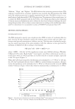
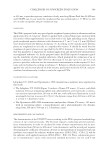
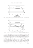
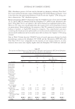
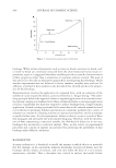
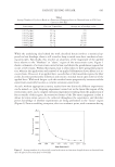
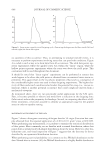
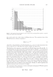
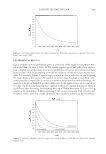
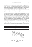
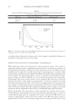
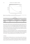
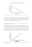
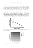
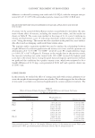
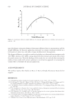
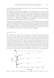
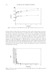
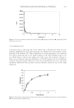
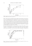
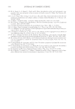
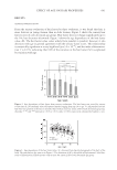
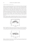
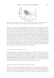
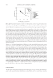
JOURNAL OF COSMETIC SCIENCE 634 Figure 7 shows the release profi les from all the samples studied, with the release percent- age of active ingredient practically reaching 100% in all cases. The release of Hydrocotyle asiatica from both formulations was similar, both in terms of total quantity released, as well as in terms of time taken for a 100% release, taking place two hours after initiation of the test. However, differences for the Aloe samples were observed, with 100% release taking place after two hours for the organogel and after four hours for the hydrogel. How- ever, we can confi rm that the release of the active principle from the excipients used in our study was appropriate, both in terms of magnitude and rate, and that the active ingredi- ents will be effective after application to the skin. The results obtained from the in vitro release model used in this study enable us to esti- mate the in vivo release behavior of the formulae tested, subject to the same parameter variables (7,13). The differences found in the time taken for the 100% release of Hydro- cotyle asiatica and the Aloe in the same media could be attributable to differences in Figure 6. Percentage of drug solution released with each type of membrane for Hydrocotyle asiatica: nylon membrane ( ) and methylcellulose membarnce ( ). Figure 7. Percentage of drug released for different formulations: Aloe hydrogel (♦), Hydrocotyle asiatica hydrogel ( ), Aloe PLO ( ), and Hydrocotile asiatica PLO (×).

HYDROGELS AND ORGANOGELS AS VEHICLES 635 physical–chemical properties such as solubility and partition coeffi cient. In addition, pluronic gel is hydrophilic whereas the PLO base is lipophilic (28). CONCLUSIONS On the basis of these results, we can confi rm that all of the formulations studied had ap- propriate organoleptic characteristics as well as a good rheological plastic behavior. Com- pression force values were low, despite the fact that the organogels presented higher viscosity. These properties give them a pleasant texture, allowing them to be spread easily over the skin. On the other hand, the in vitro release profi les reveal that the delivery of the drugs from the tested excipients is appropriate, both in magnitude and in time. REFERENCES (1) G. Yosipovitch, A. DeVore, and A. Dawn, Obesity and the skin: Skin physiology and skin manifesta- tions of obesity, J. Am. Acad. Dermatol., 56(6), 917–919 (2007). (2) G. E. Pierard, J. L. Nizet, and C. Pierard-Franchimont, Cellulite from standing fat hemiation to hypo- dermal stretch marks, Am. J. Dermatopathol., 22, 34–37 (2000). (3) L. Benelli, J. L. Berta, and C. Camistra, Endermologie: Humoral repercussions and estrogen interaction, Aesthetic Plast. Surg., 23, 312–315 (1999). (4) N. S. Sheinfeld, Obesity and dermatology, Clinics Dermatol., 22(4), 303–309 (2004). (5) L. Batllori, Aloes, Offarm., 11, 67–68 (1992). (6) R. Gampel, Propiedades y utilidades del Aloe vera, Ponencia en las Jornadas de Fitoterapia y Etnobotánica, n° 117, Madrid (2002). (7) C. A. Guzzo, G. S. Lazarus, and V. P. Perth, “Dermatological Pharmacology,” in The Pharmacological Basis of Therapeutics, 9th ed., J. G. Hardman, L. E. Limbird, P. B. Molinoff, R. W. Ruddan, and A. G. Gilman, Eds. (McGraw Hill, New York, 1996). (8) H. William and P. L. Luisi, Lecithin organogels as matrix for transdermal transport of drugs, Biochem. Biophys. Res. Commun., 177, 897–900 (1991). (9) S. Murdan, A review of pluronic lecithin organogels as a topical and transdermal drug delivery system, Hospital Pharmacist, 12, 267–270 (2005). (10) J. Franckum, D. Ramsay, N. G. Das, and S. K. Das, Pluronic lecithin organogel for local delivery of anti-infl ammatory drugs, Int. J. Pharm. Comp., 8, 101–105 (2004). (11) R. Kumar and O. P. Katare, Lecithin organogels as a potenthial phospholipid-structured system for topical drug delivery: A review, AAPS Pharm. Sci. Tech., 6(2), 298–310 (2005). (12) R. C. Wester and H. I. Maibach, Cutaneous pharmacokinetic: 10 steps to percutaneous absorption, Drug Metab. Rev., 14, 169–205 (1983). (13) I. A. Alkrad, Y. Mrestani, and R. H. Neubert, The release profi les of intact and enzymatically digested hyaluronic acid from semisolid formulations using multi-layer membrane system, Eur. J. Pharm. Bio- pharm., 56(1), 37–41 (2003). (14) R. Gupta and S. J. Flora, Effect of Centella asiatica on arsenic induced oxidative stress and metal distri- bution in rats, J. Appl. Toxicol., 26, 213–222 (2006). (15) K. Punturee, C. P. Wild, W. Kasinrerk, and U. Vinitketkumnuen, Immunomodulatory activities of Centella asiatica and Rhinacanthus nasulus, Extracts Asian Pacifi c J. Cancer Prev., 6, 396–400 (2005). (16) G. Jayashree, G. Kurup Muraleedhara, S. Sudarslal, and V. B. Jacob, Anti-oxidant acitivity of Centella asiatica on lymphoma-bearing mice, Fitoterapia, 74(5), 431–434 (2003). (17) S. Sasaki, H. Shinkai, Y. Akashi, and Y. Kishihara, Studies on the mechanism of action of asiaticoside (Madecassol®) on experimental granulation tissue and cultured fi broblasts and its clinical application in systemic scleroderma, Acta Dermatovenerolog, 52, 141–150 (1972). (18) D. Randriamampionona, B. Diallo, F. Rakotoniriana, C. Rabemanantsoa, K. Cheuk, A. M. Corbisier, J. Mahillon, S. Ratsimamanga, and M. El Jaziri, Comparative analysis of active constituents in Centella asiatica samples from Madagascar: Application for ex situ conservation and clonal propagation, Fitotera- pia, 78, 482–489 (2007).
Purchased for the exclusive use of nofirst nolast (unknown)
From: SCC Media Library & Resource Center (library.scconline.org)