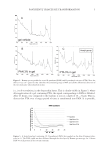
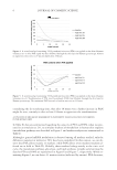
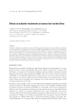
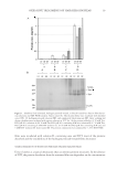
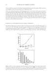
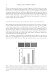
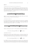
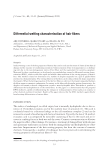
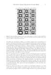
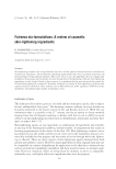
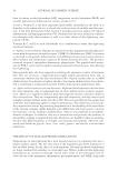
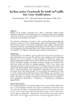
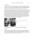
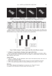
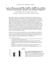
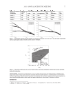
JOURNAL OF COSMETIC SCIENCE 20 of urea, while the protein dissolution from the hydrogen peroxide-treated fi lms increased only slightly as urea increased from 0 to 8 M (Figure 2A). Next, we examined the effects of hydrogen peroxide concentration on the protein dissolu- tion from the keratin fi lms (Figure 2B). The amount of proteins dissolved in solution B began to decrease when the concentration of hydrogen peroxide was more than 0.001%. Dissolved proteins from the fi lms decreased linearly when the fi lms were treated with increasing concentrations of hydrogen peroxide. The half-value was estimated to be ca. the 0.01% hydrogen peroxide solution, indicating that the hair protein fi lm was highly sensitive to the oxidative processing. MORPHOLOGY OF THE KERATIN FILMS WITH VARIOUS TREATMENTS The keratin fi lms were incubated with 1% hydrogen peroxide or the bleaching agent at 25°C for 10 min. The fi lms remained opaque irrespective of the hydrogen peroxide and the bleaching treatments (Figure 3A, panels a, b, c). However, the color was changed to the naked eye, from light brown to white, by the bleaching treatment, while the hydrogen Figure 2. Effects of urea and hydrogen peroxide on protein solubility from the hair keratin fi lms. (A) The keratin fi lms treated with distilled water ( ) and 1% hydrogen peroxide ( ) were incubated with solution A containing 0–8 M urea. The protein concentrations of the supernatants were measured. (B) The keratin fi lms were treated with 0.001–10% hydrogen peroxide and the solubility of solution B was examined.

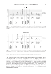
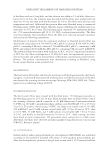
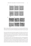
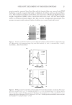
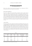
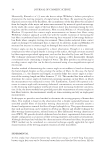
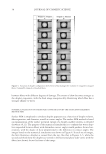
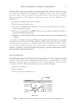
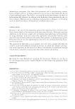
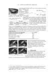
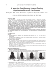
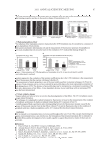
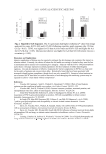
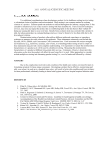
OXIDATIVE TREATMENTS OF HAIR KERATIN FILMS 21 Figure 3. Changes in appearances of the hair keratin fi lms after various treatments and SEM observations. (A) The hair keratin fi lms were incubated with distilled water (a,d), 1% hydrogen peroxide (b,e), and bleach solution (c,f) at 25°C for 10 min. The fi lms (d–f) were further incubated with solution B at 50°C for 3 hr. (B) After washing and drying, the fi lms were used for morphological observations. Bars: 20 mm (A) 10 μm (B). peroxide-treated fi lm apparently remained unchanged. These fi lms were incubated with solution B at 50°C for 3 hr, washed with distilled water, and dried. Proteins in the un- treated fi lm partly dissolved and became translucent, making the letters “keratin” under- neath the fi lm visible. On the other hand, proteins in the fi lms treated with hydrogen peroxide or the bleaching agent hardly solubilized, resulting in their clearly unchanged appearances (Figure 3A, panels d, e, f). We reported that the surfaces of hair protein fi lms prepared by pre- and post-cast methods were composed of porous structures with fi ne fi laments and particles (11,12). SEM observa- tion showed that the surfaces of hair keratin fi lms prepared by the pre-cast method are also composed of porous structures containing small particles (Figure 3B, panels a, b, c). Such small particles on the surface of the translucent fi lms incubated with solution B disap- peared, while smooth porous structures remained (Figure 3B, panel d). Urea treatment ap- parently could not affect the fi ne structures of the fi lms, even after the oxidative treatments.
Purchased for the exclusive use of nofirst nolast (unknown)
From: SCC Media Library & Resource Center (library.scconline.org)