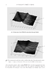
JOURNAL OF COSMETIC SCIENCE 10 topography can be achieved by applying either a contacting stylus or some sort of optical systems to the surface of the replica (2). These traditional measurement techniques applied to static silicone replicas of the skin have proved very useful in recording skin topography with satisfactory accuracy for use in analyzing skin microstructure and anisotropy. Although skin replicas have been used as a successful means to investigate the topography of the skin, there are a number of drawbacks associated with this kind of in vitro replica- tion technique. First, the silicon replica can reproduce the structure of the skin, but fails to copy the color information of the skin which is also most important in skin disease diagnosis. Second, it takes a relatively long time to collect the data, which makes it un- suitable for real applications with short time requirements, as may be the case when col- lecting data in a primary care scenario. Third, considerable operator skill is required to successfully copy the skin using the replica method. To obtain a qualifi ed replica, the sample should be taken on clean and dry skin under given specifi ed temperature and hu- midity on standardized positions of the body and with correct specifi ed mixture ratio of catalyst and paste. Finally, highly sensitive and/or damaged skin may not be durable to contact with the polymer replica materials directly. Clearly an appropriate direct observation of the skin surface is clinically desirable, par- ticularly given the aforementioned disadvantages of an indirect replica-based method. In a clinical setting, the most frequently used methods are direct manual observations with the naked eye together with the use of photography. Although these methods have been used for a long time, they tend to suffer from low accuracy and subjective judgment. Hence, more scientifi c techniques are required to be integrated in the inspection of the skin surface in vivo. Various types of video microscope have been developed to scan the skin surface, but few of them can be used with minimal patient inconvenience while in a clinical setting (3). The PRIMOS device uses a structured light source technique and has been commercialized with released specifi cations for the measurement of skin in vivo. The 3D data of the skin from this device are recovered by using a phase-shifting principle from a series of images with stripe lights projected onto the skin. It has been considered in dermatology as an ideal tool for the investigation and documentation of skin micro- structure and wrinkles (4). The temporal phase shift–based PRIMOS method is one of the most frequently used methods however its working principle, where the object is recov- ered using a serial projection of parallel black/white stripes, makes this technique unsuit- able for the recovery of heavily colored skin surface. Meanwhile, the resolution of this technique has been limited. Some experiments have pointed to an inability to record fi ne scale features as the fringe may not be able to reach deep valley features on living skin (5). In addition, the PRIMOS is a bulky device and does not lend itself to handheld use. It is almost impossible to use it for in vivo measurement of those skin lesions distributed arbi- trarily around the body. A NEW SKIN PROFILE MEASUREMENT DEVICE: SKIN ANALYZER Photometric stereo is an optical approach to recover the surface shape of an object using several images taken from the same view point but under different lighting directions and has been extensively used in industrial applications, especially for inspection of large-scale products such as tile, stones, and metal components. Because of the relative unrestricted availability of space in the design and construction of these optical systems,

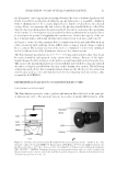
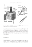
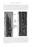
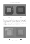
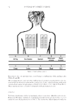
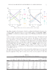
EVALUATION ON AN OPTICAL SCANNING DEVICE 11 the illuminates can be appropriately arranged around the objects without signifi cant dif- fi culty to satisfy the assumption of achieving a point light source or a parallel, collimated form of illumination (6). It is clearly impractical to handle a bulky device in a clinical setting. Hence, an ergonomically and aesthetically pleasing handheld device called Skin Analyzer has been designed and manufactured with the aim of estimating the topography of skin surface (7). Several pilot clinical studies have been undertaken by using this device to investigate the potential of applying the new device to obtain a description of the sur- face of human skin as additional information for monitoring local skin conditions (8). As Figure 1 shows, the Skin Analyzer device is fi nished in black and embedded with six surface-mounted light-emitting diodes (LEDs) and a compact digital Charge-Coupled Device camera. The internal structure of the device is confi gured to achieve the optimized results for the position of the illuminates relative to the camera system. The Skin Analyzer has dimensions of 65 * 65 * 130 mm and weighs less than 1 kg. It can be easily assembled and operated using custom-build software. Because of the light- weight design, the Skin Analyzer can be held in a single hand without touching the skin. This ensures the measurements have good repeatability and stability because any external distortion is largely excluded from the skin surface during data capture. The following evaluations on the device whose manufacturing details are described in (9) are carried out on both skin replica in vitro and skin lesion in vivo by comparing with the reference data acquired by the PRIMOS. EXPERIMENTAL EVALUATION ON SKIN PROFILE RECOVERY THE COMPARISON DATA FORMAT The Skin Analyzer provides surface gradient information directly based on the principle of photometric stereo. The gradient data are the results of partial differentiation of the Figure 1. Schematic (A) and developed (B) handheld skin lesion imaging system – known as the “Skin Analyzer” composed of a small IEEE1394 digital camera (AVT Marlin, F-046C) and a high-resolution com- pact lens (Schneider, 1.4/23 mm + extension tube), surrounded by six surface-mounted high power chip-type LEDs (NSCW455, NICHIA, Tokushima, Japan) positioned equidistantly in angle on a circle of radius 8 cm that is centered on the camera’s optical axis and lies in a plane orthogonal to this axis in line with the front of the fi rst optical element of the lens.
Purchased for the exclusive use of nofirst nolast (unknown)
From: SCC Media Library & Resource Center (library.scconline.org)