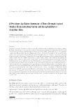
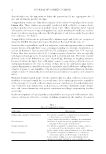
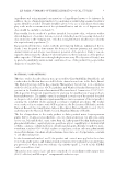
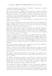
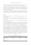
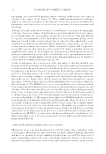
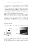
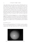
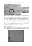
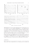
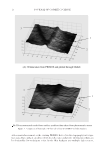
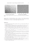
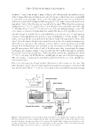
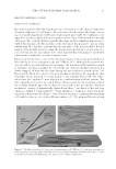
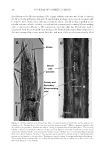
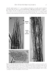
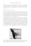
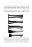
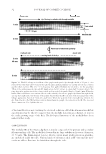
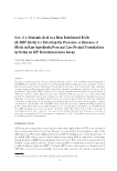
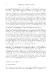
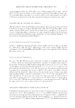
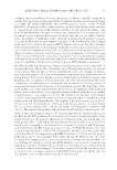
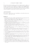
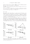
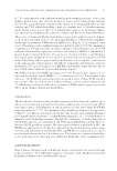
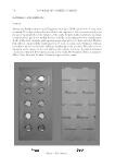
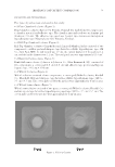
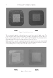
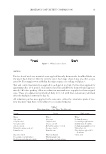
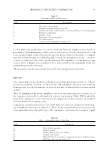
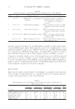
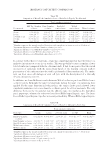
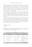
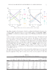
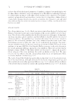
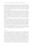
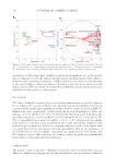
EVALUATION ON AN OPTICAL SCANNING DEVICE 15 Figure 6. Needle map s of the skin replica. The Skin Analyzer also keeps the profi le changing smoothly. Such a phenomenon can be observed from the reconstructed profi le maps in Figure 7, i.e., some small deeper holes (1, 2, 3) appear roughly recovered using the PRIMOS. This may be explained by a failure in the recovery of features with sharp edges due to the obstruction of projection light il- luminated from one direction only. The Skin Analyzer is able to take advantage of the multiple light sources to remove the presence of shadow and specularities. From the profi le maps, the edges and holes reconstructed using the photometric stereo technique appear more reliable and credible than those recovered from the PRIMOS. Figure 8 shows images rendered with a virtual light using the gradient data from both the Skin Analyzer (A) and the PRIMOS (B). It can be found that the Skin Analyzer can recover the skin, but PRIMOS failed to detect the details of live skin. The data collected by the PRIMOS on in vivo skin tend to be even less credible as the PRIMOS needs a long acquisition time and cannot compensate for object (usually human being) movement. CONCLUSION From the experimental evaluation presented in this article, it is proven that the Skin Analyzer device based on the photometric stereo technique may be more suitable than

JOURNAL OF COSMETIC SCIENCE 16 Figure 7. Comparison of skin replica 3D data taken from the PRIMOS and Skin Analyzer. either manual assessment or the existing PRIMOS device for skin topography investiga- tion tasks. Especially, it can deliver both the refl ectance and profi le information which can be obtained by few techniques so far. As the Skin Analyzer uses multiple light sources,
Purchased for the exclusive use of nofirst nolast (unknown)
From: SCC Media Library & Resource Center (library.scconline.org)