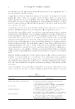
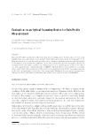
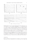
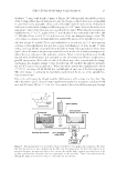
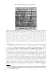
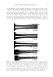
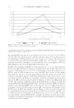
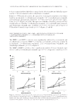
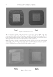
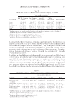
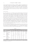
EVALUATION ON AN OPTICAL SCANNING DEVICE 11 the illuminates can be appropriately arranged around the objects without signifi cant dif- fi culty to satisfy the assumption of achieving a point light source or a parallel, collimated form of illumination (6). It is clearly impractical to handle a bulky device in a clinical setting. Hence, an ergonomically and aesthetically pleasing handheld device called Skin Analyzer has been designed and manufactured with the aim of estimating the topography of skin surface (7). Several pilot clinical studies have been undertaken by using this device to investigate the potential of applying the new device to obtain a description of the sur- face of human skin as additional information for monitoring local skin conditions (8). As Figure 1 shows, the Skin Analyzer device is fi nished in black and embedded with six surface-mounted light-emitting diodes (LEDs) and a compact digital Charge-Coupled Device camera. The internal structure of the device is confi gured to achieve the optimized results for the position of the illuminates relative to the camera system. The Skin Analyzer has dimensions of 65 * 65 * 130 mm and weighs less than 1 kg. It can be easily assembled and operated using custom-build software. Because of the light- weight design, the Skin Analyzer can be held in a single hand without touching the skin. This ensures the measurements have good repeatability and stability because any external distortion is largely excluded from the skin surface during data capture. The following evaluations on the device whose manufacturing details are described in (9) are carried out on both skin replica in vitro and skin lesion in vivo by comparing with the reference data acquired by the PRIMOS. EXPERIMENTAL EVALUATION ON SKIN PROFILE RECOVERY THE COMPARISON DATA FORMAT The Skin Analyzer provides surface gradient information directly based on the principle of photometric stereo. The gradient data are the results of partial differentiation of the Figure 1. Schematic (A) and developed (B) handheld skin lesion imaging system – known as the “Skin Analyzer” composed of a small IEEE1394 digital camera (AVT Marlin, F-046C) and a high-resolution com- pact lens (Schneider, 1.4/23 mm + extension tube), surrounded by six surface-mounted high power chip-type LEDs (NSCW455, NICHIA, Tokushima, Japan) positioned equidistantly in angle on a circle of radius 8 cm that is centered on the camera’s optical axis and lies in a plane orthogonal to this axis in line with the front of the fi rst optical element of the lens.

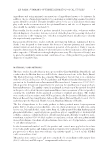
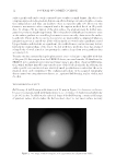
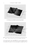
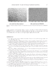
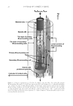
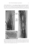
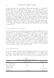
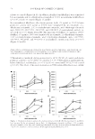
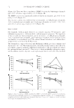
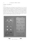
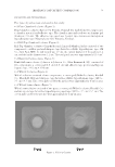
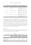
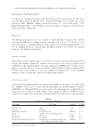
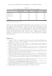
JOURNAL OF COSMETIC SCIENCE 12 surface profi le and can be easily converted into a surface normal format. An object de- scription expressed in the gradient domain can offer advantage in terms of surface orienta- tion independence and thus can facilitate object recognition tasks (10). However, this format is not intuitive when compared with a description method based on 3D profi le data. To obtain a 3D description of the skin surface, the gradient must be further inte- grated to produce a height-map format. This is found to be diffi cult and sensitive to noise as the surface gradient recovered by photometric stereo can only characterize the surface locally (11). Errors in the reconstruction process are unavoidably accumulated when an integration process is carried out. If the outliers in the recovered surface gradients caused by specularities and shadows are signifi cant, the results from reconstruction may deviate far from the original shape of the object. To deal with these problems, there has emerged a large body of work aimed at integrating the surface shape from noisy gradient data accurately (12). To make the data extracted using the photometric stereo technique comparable with that of the pure 3D data output from the PRIMOS device, we transform the 3D data from the PRIMOS into a gradient representation format using a procedure of partial differentia- tion, which will not introduce any error because of the local calculation. In addition, the surface profi le is reconstructed on a relatively small specifi ed area in order that the accu- mulated errors can be minimized. The evaluation procedure on the acquired topography data is carried out using three test objects, i.e., a painted ball bearing, replica of skin, and live skin. THE EXPERIMENTAL SUBJECTS Ball bearing. A ball bearing with diameter of 10 mm in Figure 2 is chosen as a reference because it is manufactured with high precision, i.e., tolerance of diameter and sphericity is ±0.002 inches. In addition, the spherical shape of the ball bearing contains a full range of gradient values, which makes the ball an ideal object to test most surface recovery Figure 2. One image of ball bearing able to be approximated as a Lambertian surface.
Purchased for the exclusive use of nofirst nolast (unknown)
From: SCC Media Library & Resource Center (library.scconline.org)