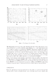
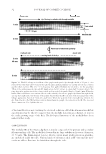
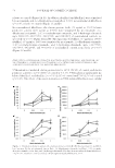
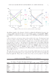
REGULATION OF EXTRACELLULAR MATRIX BY NICOTINAMIDE AND ITS DERIVATIVES 49 (1–4,8,27). One hundred microliter aliquots of media or cells from each sample, or respective standards were added to independent wells of 96 well plates for 24 h. The wells were blocked with bovine serum albumin, and then incubated with respective antibod- ies (Elastin Products Co.) for 1 h. The plates were washed with wash buffer, incubated with respective secondary antibodies linked to peroxidase for 1 h, washed, and subse- quently incubated with peroxidase substrate until color development, which was mea- sured spectrophotometrically at 405 nm and quantitated from standard curves. ELASTIN PROMOTER ACTIVITY Fibroblasts were cotransfected with elastin promoter-fi refl y luciferase (Pgl4 vector) and TK-Renilla luciferase plasmids (for normalization of transfection effi ciency) using Escort (Sigma) for 24 h before dosing with or without UVA-radiation ± niacin derivatives for 24 h (2,4,8). The cells were measured for luminescence from fi refl y or renilla luciferase with specifi c substrates and quantitated using recombinant luciferase as standard (Promega). MMP-1, -9, AND -9, AND ELASTASE ACTIVITIES The inhibition of ECM proteolytic enzymes (MMP-1, MMP-2, MMP-3, Elastase) (Biomol, Torrance, CA Enzo Life Sciences, Farmingdale, NY Elastin products Co.) was per- formed as previously reported (1,3). Each of the enzymes at optimal concentration was incubated with the niacin derivatives at 0%, 0.01%, 0.1%, or 1% for 10 min followed by the addition of its respective substrate (Bachem). The reaction kinetics were measured fl ourometrically (355 excitation/450 emission) every 10 min for a total of 60 min. The initial reading (0 time) was subtracted from the fi nal reading (60 min) and the data con- verted to percentage of control. DATA ANALYSIS The data were analyzed for signifi cant difference by analysis of variance and student t-tests at 95% confi dence interval. The effects of UVA radiation on dermal fi broblasts were analyzed relative to nonirradiated control cells. The effects of the niacin derivatives on nonirradiated cells were analyzed relative to nonirradiated cells (control). The effects of each of the niacin derivatives on UVA-radiated fi broblasts were analyzed relative to UVA radiation effect alone (UVA-radiated respective control). The direct MMP or elastase in- hibitory activity of the niacin derivatives was analyzed relative to control. RESULTS STIMULATION OF EXPRESSION OF ELASTIN, ELASTIN PROMOTER, FIBRILLIN-1, AND FIBRILLIN-2 BY NICOTINAMIDE, 2,6-DIHYDROXYNICOTINAMIDE, 2,4,5,6-TETRAHYDROXYNICOTINAMIDE, AND 3-HYDROXYPICOLINAMIDE IN NONIRRADIATED FIBROBLASTS Niacin or its derivatives stimulated the expression of elastin and fi brillin in dermal fi bro- blasts. The niacin derivatives signifi cantly stimulated elastin expression (protein and pro- moter) and fi brillin-1 and fi brillin-2 at 0.1% and 1% in nonirradiated fi broblasts ( p 0.05,

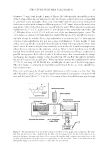
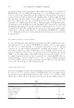
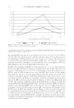
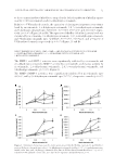
JOURNAL OF COSMETIC SCIENCE 50 relative to control) (Figure 1A–D). In addition, fi brillin-1 and fi brillin-2 were stimulated by nicotinamide and 2,6-dihydroxynicotinamide at 0.01% in nonirradiated fi broblasts ( p 0.05, relative to control) (Figure 1C and D). In nonirradiated fi broblasts, the elastin protein levels (70 ng/ml as 100%)/elastin promoter activity (400 pg/ml as 100%) were stimulated by nicotinamide, 2,6- dihydroxynicotinamide, 2,4,5,6-tetrahydroxynicotinamide, and 3-hydroxypicolinamide upto 308/222%, 413/719%, 243/358%, and 224/183% of nonirradiated controls, re- spectively ( p 0.05) (Figure 1A and B). The expression of fi brillin-1 (0.5 μg/ml as 100%)/ fi brillin-2 (35 ng/ml as 100%) was stimulated by nicotinamide, 2,6-dihydroxynicotinamide, 2,4,5,6-tetrahydroxynicotinamide, and 3-hydroxypicolinamide upto 614/578%, 601/533%, 402/234%, and 336/290% of nonirradiated controls, respectively ( p 0.05) (Figure 1C and D). STIMULATION OF EXPRESSION OF ELASTIN, ELASTIN PROMOTER, FIBRILLIN-1, AND FIBRILLIN-2 BY NICOTINAMIDE, 2,6-DIHYDROXYNICOTINAMIDE, 2,4,5,6-TETRAHYDROXYNICOTINAMIDE, AND 3-HYDROXYPICOLINAMIDE IN UVA RADIATED FIBROBLASTS UVA-radiation stimulated elastin protein level to 185% (±14%) of control, and elastin promoter activity to 125% (±8%) of control (p 0.05). UVA-radiation signifi cantly in- hibited fi brillin-1 and fi brillin-2 to 65% (±4%) of control and 73% (±7%) of control ( p 0.05). The effects of the niacin derivatives on UVA-radiated fi broblasts were similar Figure 1. Stimulation of elastin protein (A), elastin promoter activity (B), fi brillin-1 protein (C), and fi bril- lin-2 protein (D) by nicotinamide (green line), 2,6-dihydroxynicotinamide (red line), 2,4,5,6-tetrahy- droxynicotinamide (violet line), and 3-hydroxypicolinamide (blue line) in non-irradiated dermal fi broblasts * = p 0.05, relative to control, error bars (A–D) represent standard deviation, n = 4.
Purchased for the exclusive use of nofirst nolast (unknown)
From: SCC Media Library & Resource Center (library.scconline.org)