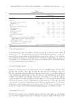
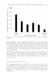
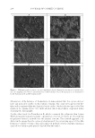
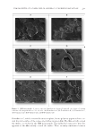
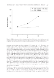
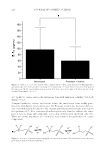
UNDERSTANDING SOLAR SKIN ELASTOSIS 177 REAL-TIME PCR FOR ANALYSIS OF ELASTIN AND LOXL1 EXPRESSION LEVELS Cell culture. Fibroblasts (63-year-old donor) were cultured until confl uence. They were then exposed to ultraviolet A (UVA) (7.5 J/cm2) and cultured for 24 h. LOXL1 and elas- tin expression was evaluated by quantitative - reverse transcription - polymerase chain reaction (q-RT-PCR). In the second experiment, fi broblasts (63-year-old donor) were cultured until confl uence. Three different conditions have been used. The control culture was maintained for 24 h and then exposed to UVA (7.5 J/cm2). The culture was maintained for 16 h. In the post irradiation condition, the Hamamelis extract was added just after the irradiation, and maintained for 16 h. In the pre- and postirradiation conditions, the specifi c Hamamelis extract was added in the culture at confl uence and for 24 h. Then the culture was exposed to UVA (7.5 J/cm2) and the Hamamelis extract was added for additional 16 h. Assay method. The cells were washed with phosphate buffer solution (PBS) and then total RNA was extracted (Spin Vacuum Total RNA Isolation System Z3500, Promega, Char- bonniere les bains, France). Total RNA content and quality were evaluated by measuring optical density at 260 and 280 nm. Real-time RT-PCR was performed using iScript one-Step Real-time reverse transcription-polymerase chain reaction (qRT-PCR) Kit with SYBRgreen (Bio-Rad, Marnes-la-Coquette, France). The following primers were used for PCR: For LOXL1. Forward 5′-GACTTCGGCAACCTCAAGC-3′ Reverse 5′-TGTTGCAGAAACGTAGCGAC-3′ For elastin. Forward 5′-GTGTATACCCAGGTGGCGTG-3′ Reverse 5′-CGAACTTTGCTGCTGCTTTAG-3′ All primers were in separate zone of the exon. Amplifi cation was performed with 40 cycles, measuring the fl uorescence at the end of each cycle. The comparative Ct method (f¢Ct) was used for relative comparison. Real-time PCR experiments were calibrated with actin as the housekeeping gene. As negative controls, samples without RNA were used in the same conditions. Results were expressed related to control (untreated irradiated cells) and normalized to actin. Results and statistics. The results are expressed as percentage compared with untreated con- trol and then expressed as mean ± standard deviation (SD) from nine replicates. The statis- tical analysis was carried out using a Student t-test. IMMUNOHISTOCHEMISTRY: ELAFIN IN NORMAL HUMAN BIOPSIES EXPOSED TO UVA Cell culture. Human biopsies (abdominal part) from a 27-year-old donor were cultured in a specifi c defi ned medium at 37°C, with 5% CO2, for 10 d. The biopsies were either ir- radiated or not with UVA at 5 J/cm2 during a period of 10 d. UVA were applied from day 2 with and without our specifi c Hamamelis extract at 0.5%, every day in a topical or systemic manner. Assay method. Samples were fi xed in a formalin solution and they were then dehy- drated and embedded in paraffi n. Seven-micrometer sections were deparaffi nized.

JOURNAL OF COSMETIC SCIENCE 178 The antihuman elafi n antibody (Abcam) was used at a concentration of 1/200 for one night at room temperature and amplifi ed with streptavidin/biotin. The technique involves three layers. The fi rst layer is an unlabeled primary antibody. The second layer is a biotinylated secondary antibody. The third layer is a complex of streptavidin–biotin peroxidase. The peroxidase (CliniSciences, Nanterre, France) produces purple-colored end products. They were observed using an optical microscope (Leica DMLB, Nanterre, France). Quantifi cation. The staining was detected with acquisition of a threshold that allowed selecting the staining in the interest area. The surfaces were then measured. The data were exported as values in an Excel fi le. Results and statistics. The quantifi cation of each band was relative to actin. The results were expressed in the protein percentage compared with the untreated control at 100%. Statistics were analyzed using a Mann–Whitney nonparametric test. IN VIVO CLINICAL TESTS Study design. The clinical study was carried out as a placebo-controlled double-blind randomized split-face study under dermatological control. The effi cacy of the formula containing Hamamelis at 1% was compared with the baseline (before treatment, D0) and to the other half-face treated with placebo. The study was conducted during a period of 56 d with check points at D0, D28, and D56 for the antiwrinkle effect, and during a period of 84 d with check points at D0 and D84 for the fi rming effect. This study was carried out in France from March to June 2013. Inclusion criteria. The study was performed on Caucasian female volunteers aged from 52 to 76 year, displaying wrinkles and fi ne lines on the crow’s feet of average to strong intensity and saggy skin on the face. Application modality. The products (containing Hamamelis at 1% or placebo formula) were applied by the volunteers twice a day, on each half face for 84 d. The application was carried out by the volunteers, by circular massages until complete penetration, especially on the crow’s feet area and the cheek. METHOD OF EVALUATION Measurement of skin roughness by fringe projection. Images were acquired on the crow’s feet area (surface of 12 cm2) via digital video camera coupled to a fringe projection system (Dermatop™ system, Breuckam, Meersburg, Germany, Eotech, Marcoussis, France) at D0, D28, and D56. ST and Stm parameters were calculated as follows (TopoSurf and Optocat analysis systems): - ST: maximum amplitude of the relief (mm). A decrease means a reduction of the main wrinkles. - Stm: mean difference b etween peaks and valleys (mm). A decrease means a smoothing of the studied surface. Measurement of the fi rmness of the skin with Dynaskin®. Dynaskin® (Orion Concept, Tours, France) is an add-on to the DermaTop™ system, which permits to evaluate the
Purchased for the exclusive use of nofirst nolast (unknown)
From: SCC Media Library & Resource Center (library.scconline.org)