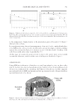
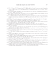
HAIR SHAFT FORMATION AND MITOCHONDRIAL BIOENERGETICS 325 BIOLOGY: THE HAIR FOLLICLE AND THE PROLIFERATION STAGE Hair follicles are complex skin-associated organs within the dermis/epidermis (Figure 1), where each “pilosebaceous” or “follicular” unit comprises the hair synthesis machinery, the sebaceous (oil) gland, the apocrine (sweat) gland, and an arrector pili muscle (15,16). Each follicle consists of both epithelial and mesenchymal components: the outer root sheath (ORS), inner root sheath (IRS), and germinative matrix (GM) are epithelial in derivation, whereas the dermal papilla (DP) and encapsulating connective tissue sur- rounding the follicle known as the dermal sheath (DS) are mesenchymally derived (17,18). Importantly for full understanding of follicle function, the dermal components are vascu- larized and enervated. On human scalp, most of the cells within the GM eventually form the cortex of the hair shaft. Although these structures are derived from the same source, the division of the scientifi c bodies researching hair production often has differing lexicons and confusing nomenclatures. As this is an attempt to bring together these disparate fi elds, societies, and institutions, we will attempt to defi ne each group’s terminology. Throughout this review, where we see confusion or duplication, we will revert to the nomenclature defi ned in Figure 1. The follicle bulb creates hair shaft and other support layers similar to how the epidermis creates the stratum corneum: basal cells progressively differentiate, die, and cornify into the dead outer protective layer (20). In the follicle bulb, a basal epidermal layer attached to a basement membrane surrounding the DP rapidly divides and produces a GM. It is believed that each cell lining along the DP membrane is fully fated to a specifi c lineage to create a specifi c hair follicle layer. Maintenance of a healthy follicle is fundamentally Figure 1 . Nomenclature of the key structures of the human hair follicle bulb. Reprinted by permission from Springer Nature: Introduction to hair development by D. Harland, Copyright 2018 (19).

JOURNAL OF COSMETIC SCIENCE 326 dependent on epithelial–mesenchymal interactions across this complex basement mem- brane and how each follicular compartment communicates with the others (21). The DP is quiescent, essentially never containing mitotic activity and having relatively low met- abolic activity but likely controls the number, size, and fate of all the GM cells. The DP thus determines the fi ber size, content, and hence fi nal cosmetic properties (17,22). As GM cells cease division and migrate above the Auber’s line, differentiation begins: they pack closely, compress and align along the soon to be fi ber axis (23,24). In some hairs, other GM cells migrate to a centrally located medulla, whereas in all hairs, the cortex is surrounded by cells that will become the hair shaft cuticle. The outer follicular layers that provide structure and support during shaft growth also originate in the GM and migrate and differentiate similarly to the shaft cell lines. The IRS differentiates into three cell lines named medially to laterally: the IRS cuticle, Huxley’s layer, and Henle’s layer. Adjacent to Henle’s layer is a distinct layer of fl attened, elongated cells termed the “companion layer.” The companion layer is closely associated with both Henle’s and Huxley’s layers through numerous direct connections, leading to speculation that the companion layer nourishes the IRS and may even guide its differen- tiation and eventual destruction in the follicle lumen (25–27). The aforementioned IRS layers surround and move with the hair shaft – progressing outward to the skin surface and then disintegrate into the follicle infundibulum (where the follicle reaches the skin surface). The companion layer is hence most likely the “shear plane” between the outwardly moving hair shaft and its associated structures, and the stationary, ORS. The ORS resides outside the companion layer and consists of characteristic cuboidal cells that thicken from one to multiple layers as the follicle extends distally. At the proximal end, the ORS is contiguous with GM cells lining the lower half of the DP. At the distal end, the ORS is contiguous with the interfollicular epidermis. Interestingly, these cells are competent to regenerate a stratifi ed, fully differentiated interfollicular epithelium in vitro (28). ORS cells express keratins, keratin-associated proteins (KAPs), adhesion mol- ecules, and signaling molecules uniquely distinguishing them from interfollicular epider- mis (17,29,30). The human stem cell compartment is located near the attachment of the arrector pili muscle. In humans, it is important to note that the “stem cell” cluster of the ORS does not form a visible “bulge” as it does in murine follicles. Finally, the entire epi- dermal follicle apparatus is encased in a tough, mesoderm-derived connective tissue which forms a robust DS, from which it is separated by a thick basement membrane. CHANGES IN BIOLOGY LEAD TO A DIFFERENT PRODUCT There are numerous examples of biology-mediated alterations in hair follicles that result in direct, if diffi cult to explain, alterations in hair shaft properties. For example, monile- thrix (OMIM #158000) was described in 1879 (31) as a genetic hair disease where the hair shaft has a beaded appearance (32). The between-bead regions are breakage prone and lead to fragile hair and, hence, alopecia. Monilethrix has unknown and complex genetics and clinical overlap with other hair disorders such as localized autosomal recessive hypo- trichosis (LAH). The most of the data suggest that the disease may be caused by muta- tions in hair cortex keratin or KAP genes and/or the cell–cell contact–associated gene desmoglein 4 (32–37). The obvious hereditary nature of monilethrix, the obvious hair phenotype, and the lack of a clear cause emphasizes the complex nature of hair shaft biology.
Purchased for the exclusive use of nofirst nolast (unknown)
From: SCC Media Library & Resource Center (library.scconline.org)