HAIR SHAFT FORMATION AND MITOCHONDRIAL BIOENERGETICS 327 Many of the early hair development events we described previously and which occur in the lower bulb are fi rmly based in the fi eld of cell biology. However, many of the traits, especially those that are functional only in the dead hair, must undergo fi nal transforma- tive processes within an environment in which conventional cell biology processes may hinder that function. For example, the fi nal organization of the disulfi de network within macrofi brils occurs in a densely packed molecular environment in which enzymes and chaperone molecules cannot easily function. These stages of development fall within the fi eld of protein chemistry. Explaining the full development of normal hair, or the etiology of monilethrix, requires an interdisciplinary effort, with fi nal hair functional phenotype falling into the area of physical material science. CHEMISTRY: CELL LINE–SPECIFIC MODES OF MATURATION A feature of all cell lines in the developing fi ber, IRS, and companion layer is that the cyto- plasm is packed with free-fl oating ribosomes (38,39). Protein expression directly into the cytoplasm without modifi cation or packaging is important to the interactions that lead to structure development. Production and assembly of structural proteins must also continue within cortical and other cell lines even at late stages of fi ber development, distal of the bulb, where the cytoplasmic space is becoming crowded, the chemical environment becomes increasingly oxidative, and the organelles are being actively degraded (40). As each cell line moves up the bulb distal to its origin at the edge of the DP basement membrane, it progresses down a specifi c path of differentiation, keratinization, cornifi cation, and death. Four distinct patterns of maturation occur, and within these, each cell line has its own specifi c timing and features. Type I maturation includes the ORS, companion, Henle’s and Huxley’s layers, and the IRS cuticle, and all share sudden, demarcated cornifi cation (41) occurring within one cell length (42–44), similar to generation of cornifi ed cell envelopes in skin (45). Type II maturation occurs in the cuticle and is a prolonged multistage keratinization, which is far from understood but involves integration of nonfi lament-forming keratin and KAPs into amorphous layers, with introduction of enzymatically induced isopeptide bonds and nonenzymatic disulfi de bonds (46). Type III maturation takes place in the medulla. The medullary core of some hairs corni- fi es relatively suddenly, and the process also involves transglutaminase, nonfi brillar keratin, and trichohyalin. However, the mechanism differs from that in the root sheath (14,47). Type IV cortical maturation is gradual, nonenzymatic, involves large-scale protein con- formational changes, and cumulates in a relatively sudden cornifi cation event, which, along with the medulla and cuticle, appears to be a dewatering process in which the fi ber shrinks about 25% in diameter (48–50). ORWIN’S TRANSITION MARKS A KEY DEVELOPMENTAL STAGE AT THE TOP OF THE BULB In the lower follicle bulb, keratinocyte division stops abruptly before the cells reach the apex of the DP, and this threshold, where differentiation replaces proliferation, is

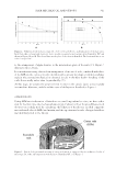
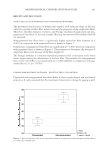
JOURNAL OF COSMETIC SCIENCE 328 known as the Auber’s critical level (51,52), or the Auber’s line, after its originator (48). Biologically driven differentiation processes dominate the remainder of the bulb, including significant cell reshaping (24) and expression of the first waves of keratins and KAPs (30). The top of the bulb is another signifi cant developmental transition. By the top of the bulb, IRS and fi ber cell nuclei have lost transcriptional function and cell shaping is complete. Around this point, high-energy metabolic events including a dramatic plume of reactive oxygen species called the “ring of fi re” is observed (53) and Henle’s layer cornifi es via a rapid enzymatic process (42,54). In the restricted tube formed by the cornifi ed Henle’s cells, with the fi ber shape fi nalized and mitochondria degrading (40), it is likely that the physical and chemical environment begins to transform into one that may be considered cytotoxic and within which maturation is driven largely by chemical processes (55). We defi ne “Orwin’s transition” as the crossover from where development is primarily driven by biology in a slightly reducing environment to one driven primarily by protein chemistry and thermodynamics in a chemically oxidizing environment. Orwin’s transi- tion crosses the follicle at the point at which Henle’s layer hardens, and in the scheme developed by Orwin, this is the border of zones B and C (19,39) (Figure 1). Distal of Orwin’s transition, macrofi brils continue to grow, but there are distinct signs of gradual keratinization in terms of protein changes (56), increasing stiffness (57), affi nity to electron microscopy stains (56) and fi lament alignment, and conformational change (57,58). Also distal of Orwin’s transition, the cytoplasmic environment is hypothesized to become chemically oxidizing, possibly because of the release of superoxide from de- grading mitochondria (40,53). The oxidizing environment encourages disulfi de bond formation and reduction in IF diameter to about 7 nm (59–62). To date, fi nal cornifi ca- tion is a process that is still not well understood (Figure 2). Figure 2. Compartmentation of hair follicle metabolism. Red, active mitochondria via tetramethylrhodamine (TMRM) green, ROS via 2′,7′–dichlorofl uorescin diacetate (DCF). The fi gure shows red proliferative matrix, weak mitochondrial activity during differentiation, and the ROS generation of the “ring of fi re” in the upper bulb of a viable, whole human hair follicle.
Purchased for the exclusive use of nofirst nolast (unknown)
From: SCC Media Library & Resource Center (library.scconline.org)