INTERNAL LIPIDS IN HAIR HEALTH 349 at 65°C with the hair and then combined and the dried residue redisolved in a second solvent (mobile phase for the SFC-MS-MS and BSTFA derivatizing reagent for the GC). Last, the bound lipids were removed with a potassium hydroxide (KOH) extraction. Hair from the free lipid extraction was digested with 50% KOH:methanol solution for 30 min at 65°C and then neutralized with 3N HCl. A fi nal extraction with chloroform:methanol solution was performed two times on the remaining hair then concentrated and made up in a second solvent. Lipids were separated using super critical fl uid chromatography, with a normal phase silica column that separated each chemical class as a group by polarity. The mobile phase was carbon dioxide with methanol with formic acid and ammonia formate modifi er. The lipids were detected with a triple quadrupole linear ion trap mass spectrometer, with multiple reaction monitoring, using atmospheric pressure ionization (APCI). Stable iso- tope-labelled internal standards for each chemical class were also used. Cetyl and stearyl alcohol were quantifi ed by gas chromatography with fl ame ionization detection using a polydimethylsiloxane capillary column with hydrogen mobile phase. Nonadecanoic acid and eicosanoic acid were used as internal standards. OPTICAL MICROSCOPY Hair samples were incubated in a solution of Nile Red (2 mg/ml) for 24 h at 37°C, before short acetone washing to remove external stain. Briefl y, hairs were cut with a sharp razor blade perpendicular to their longitudinal axis into approximately 3-cm sections. To allow cross-sectional imaging, hairs were placed in a glass capillary needle inside a 3D printed stand, with a central hole to contain the capillary tube. This assembly was then placed in a glass-bottomed dish. Stained hairs were imaged using a Zeiss 880 Airyscan Laser Scan- ning Confocal Microscope with a 1.4NA 63x oil immersion objective (Carl Zeiss Microscopy GmbH, Germany). During imaging, the 514-nm laser line was used to excite the Nile Red stain. LIPID PEROXIDE (LPO) MEASUREMENTS Hair samples were equilibrated in a 20% RH constant humidity chamber overnight. For each sample, ~0.1 g of hair was cut in 20–40-mm segments into vials (n = 4). This hair was sonicated for 2 h in a 2:1 chloroform:methanol solvent mix and then evaporated to dryness. The extract was reconstituted in isopropyl alcohol and then tested with a commercial LPO kit (KAMIYA Biomedical Company, Seattle, WA, http://kamiyabiomedical.com/). GEL NETWORK FORMULATIONS The gel network was made with 11% SLE1S surfactant, 8% stearyl alcohol, 4% cetyl alco- hol, and 77% deionized water, where all ingredients were fi rst heated together to a tem- perature between 75°C and 90°C and then cooled to room temperature. This process formed the gel premix. A small amount of this premix, 2.3% FaOH by weight of the fi nal shampoo, was then dispersed in a typical shampoo containing other benefi cial agents, such as surfactants, silicones, and polymers.

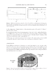
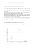
JOURNAL OF COSMETIC SCIENCE 350 HAIR FATIGUE MEASUREMENTS Hair treatments were performed on 4 g 8″ chemically treated hair switches sourced from International Hair Importers & Products, Inc. Fibers were cut from the middle of the tress and ends crimped at 30 mm using a Dia-stron Auto-Assembly System (AAS 1600) (Andover, Hampshire, UK). The average cross-sectional area along each fi ber was analyzed using a Dia-Stron Fiber Dimensional Analysis System (FDAS 770), which incorporates a Mitutoyo laser micrometer (LSM-6200) (Malborough, MA). The average cross-sectional area was calculated from three diameter measurement points along each 30-mm crimped fi ber. The average cross-sectional values for each of the fi bers were then used to set the Dia-Stron Cyclic Tester (CYC801) in controlled stress mode of 0.014 g/μm2 and rate of 40 mm/s. Environmental testing conditions were set at 50% RH and 23°C. Data were analyzed by Weibull and Kaplan–Meier statistical tools (JMP Pro 12.1.0, SAS, Cary, NC). Fibers with break cycles less than 10 were omitted from the analysis because of premature breakage. RESULTS AND DISCUSSION As we consider internal structural lipids one important question is how to quantify their levels in hair. There have been several reports in the literature (4,5) but often the data is confl icting, due in part to inherent variability in hair and in part to different extraction methods used. In this work, we developed a method using a three-stage extraction. Stage one is a hexane extraction designed to remove any surface lipids, stage two is extraction using chloroform method to extract internal unbound lipids and stage three is a hydroly- sis with KOH/methanol followed by an extraction with chloroform:methanol to extract the remaining lipids. The original intent was to use the second extraction to access un- bound lipids and the third to access bound lipids but this proved not to be possible. In- stead, 18-MEA was used as a marker for bound lipids and extraction conditions were optimized to give readily extractable unbound lipids in the second extraction and the remainder in the third extraction. Figure 1 shows data from virgin blended hair as sup- plied by International Hair Importers. Most lipids are saturated and unsaturated fatty acids with C16 and C18 chain lengths predominating, but cholesterols and ceramides were also measured. These results are comparable with those published by Masukawa et al. (6) in 2005, where a similar extraction protocol was used. These lipids are typically visualized using transmission electron microscopy but in this work confocal fl uorescence optical Fi gure 1. Internal structural lipids in hair.
Purchased for the exclusive use of nofirst nolast (unknown)
From: SCC Media Library & Resource Center (library.scconline.org)