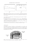
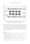
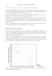
INTERNAL LIPIDS IN HAIR HEALTH 351 microscopy was used to avoid sampling artefacts. Hair was treated with Nile Red and then imaged via confocal fl uorescence microscopy in a glass capillary tube that was held vertically to enable imaging of hair cross sections in the x–y plane to give optimal spatial resolution. Nile Red only fl uoresces when associated with lipid structures and in Figure 2 this provides contrast for the hair images. Lipids are seen in the cuticle CMC and cortex CMC and highlights that these structures form a continuous network throughout hair. Areas of more intense contrast are seen in the medulla and in nuclear remnants, indicat- ing high lipid concentrations in these regions. Studies have measured changes in structural internal lipids, especially after repeated washing or coloring (3). Figure 3 shows loss of internal lipids from root to tip. Table I shows loss of internal lipids after 100 washes with a clarifying shampoo for both uncolored virgin hair and hair that had undergone fi ve cycles of coloring with an ammonia/hydrogen peroxide colorant, with fi ve wash cycles in between each coloring. Both coloring and washing remove lipids from hair and especially fatty acids. Coloring also signifi cantly decreases unsaturated fatty acid levels (~40%). It is proposed that the loss of these un- saturated lipids during coloring is because of the formation of LPOs, which can either remain in hair or undergo further oxidation, e.g. in the presence of redox metals, such as copper, to form reactive oxygen species (ROS)—see Figure 4. Experiments were carried out with eight single source-untreated virgin ponytails, where initial LPO measurements were compared with LPO levels after treating, with an ammonia/hydrogen peroxide (4.5%) colorant with very low oxidative dye levels for 30 min. All the ponytails were 25–30 cm long, Caucasian hair that had not previously been colored, and all were ap- proximately the same color (23–25 L units). To measure LPOs, hair was extracted with chloroform/methanol at room temperature and the extract measured using a LPO kit from KAMIYA Biomedical Company. In the presence of hemoglobin, LPOs are re- duced to hydroxyl derivatives (lipid alcohols) and the 10-N-methylcarbonyl-3,7- bis(dimethylamino)phenothiazine chromogen is oxidatively cleaved to form methylene blue in an equal molar reaction. LPOs are then quantitated by colorimetrically measur- ing methylene blue at 675 nm. Figure 5 shows initial LPO data from starting substrates Fig ure 2. Confocal fl uorescence microscopy image of hair stained with Nile Red dye.

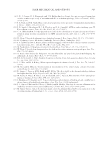
JOURNAL OF COSMETIC SCIENCE 352 and LPO levels after a 30 min colorant. LPOs were measured in almost all the starting ponytails but levels varied between ponytails. It is predicted that UV light can also form LPOs (see additional evidence below), so it is reasonable to assume these LPOs have formed from previous UV exposure by the women who donated the ponytails and may explain why levels vary from person to person. There is an increase in LPOs for all the ponytails tested apart from #1 (which had very low starting levels), providing evidence that this is a plausible mechanism for loss of unsaturated lipids after coloring. This loss is demonstrated in Figure 6, where three additional ponytails were assessed for unsatu- rated fatty acid levels after a 30-min coloring treatment. All three ponytails show a signifi cant loss of unsaturated fatty acids after treatment. The same eight ponytails were also exposed to UV irradiation that mimicked sunlight for 20 h Figure 7 shows change on LPO levels for each of the ponytails with some LPO levels increasing and some decreasing. In this case, these data illustrate that LPOs can not only be formed via UV oxidation but also be destroyed by UV oxidation. The perhydroxyl bond O-OH can be broken directly via UV absorption or more likely it reacts with other ROS formed during UV exposure. One pathway for subsequent reactivity of LPOs is shown in Figure 4 where redox metals, such as copper, can react with organic peroxides to form highly reactive alkoxyl radicals, which will react further with lipids and protein structures. Previously published work has shown such mechanisms occur with hair dosed with high versus low copper levels, with added copper and UV exposure measured LPO levels are lower than with low copper levels (7). The importance of internal structural lipids for the structural integrity of hair has been demonstrated by McMullen et al. (8), who showed removing lipids via solvent extraction caused differences in moisture management, interaction with cationic polymers and sur- factants, and impact on style hold. Fatigue measurements using fi xed weights showed a Table I L oss of lipids with washing (100 cycles) Hair treatment Sat. fatty acids (μg/g) Unsat. fatty acids (μg/g) Wax esters (μg/g) Ceramides (μg/g) Chol. + Chol. sulfate (μg/g) Total (μg/g) Vigin 8,391 ± 779 4,522 ± 252 932 ± 50 283 ± 8 1,187 ± 150 15,308 ± 1,134 +100 Wash 7,204 ± 429 2,903 ± 140 323 ± 14 293 ± 16 994 ± 70 11,727 ± 548 5 × Colored 7,312 ± 234 2,815 ± 151 404 ± 56 277 ± 19 1,108 ± 84 11,934 ± 340 +100 Wash 6,382 ± 884 2,215 ± 168 269 ± 13 269 ± 10 1,027 ± 38 10,195 ± 977 Figu re 3. Lipid loss from root to tip.
Purchased for the exclusive use of nofirst nolast (unknown)
From: SCC Media Library & Resource Center (library.scconline.org)