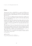
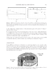
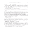
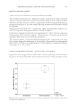
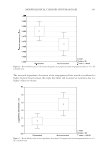
JOURNAL OF COSMETIC SCIENCE 358 by the manufacturers as being UV protectants. Hair fi bers were treated with materials of interest, exposed to a controlled amount of UV, and analyzed using Raman spectroscopy. The Atlas Suntest XXL+ (Atlas Material Testing Solutions, Mount Prospect, IL) with daylight fi lters gave UV exposure from 300 to 400 nm. The irradiance was set at 62 W/m2, which gave a total energy of 5.4 MJ/m2 over the course of 24 h. This exposure level was in line with Signori’s low UV exposure level: a few hours per day over the course of about 1 mo (1). The black standard temperature, the highest temperature the sample surfaces could reach, was set at 65°C. The relative humidity was fi xed at 55%. Raman spectra were collected using a XPlora Plus spectrometer (Horiba Scientifi c, Piscataway, NJ). The use of unpigmented hair eliminated fl uorescence and required minimal sample preparation. Raman provided a direct measure of disulfi de content and good spatial resolution with a spot size of approximately 0.5 μm. At least three hair fi bers for each treatment were fi xed to aluminum foil–covered microscope slides, as shown in Figure 1. Spectra from 200 to 3,200 1/cm were taken starting at the surface of the hair fi ber and into the hair fi ber at 0.5 μm intervals to 3 μm toward the fi ber center, giving information on the protein structure of the cuticle. These spectra were averaged, and at least three hair fi bers for each treatment were analyzed. The 785-nm laser was used at full power (100 mW at the laser source) through a ×100 objective, and 1,200 g/mm with 10-s acquisition time and three accumulations gave good signal to noise. The spectra were baselined using linear chords connected to the spectra. Spectra were normalized to peaks that did not show sig- nifi cant change upon the UV exposure used in this study: Phe at 1,003 1/cm, CH2 bending at 1,448 1/cm, amide I at 1,655 1/cm, and CH signatures at approximately 2,920 1/cm. Raman data can provide quantitative information two approaches are shown in Figure 2. The double-cursor approach to extracting data is favorable because it requires the least amount of data manipulation, but this technique cannot resolve overlapping peaks. Gaussian/Lorentzian peak fi tting allows for peak deconvolution but involves the inherent uncertainty of mathematical peak fi tting routines. Prior knowledge of anticipated peak posi- tions and reasonable initial parameters prove helpful in using this approach to peak fi tting. The Raman peaks for the different conformations of the disulfi de bond appear at different wavenumbers (9). The disulfi de peak can be deconvoluted using Gaussian/Lorentzian peak fi tting. However, for simplicity in this work due to the presence of treatment materials in addition to hair fi bers, the disulfi de peak at about 505–511 1/cm was chosen as representative of the hair fi ber disulfi de content. Gaussian/Lorentzian peak fi tting was Figure 1. Example microscope slide with unpigmented hair fi bers attached and hair fi ber schematic with Raman spectrum acquisition position indicator.

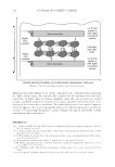
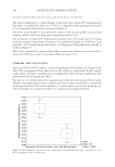
INVESTIGATING PREVENTION OF UV DAMAGE 359 crucial for understanding the potential protective effects of treatment materials A–F on hair fi ber protein structure because these materials have Raman spectra of their own. An example of peak fi tting for 440–600 1/cm for a Material B–treated, irradiated hair fi ber is shown in Figure 3. Material B has a strong peak near 488 1/cm which overlaps with the disulfi de signature from the hair fi ber. Peak fi tting was important for separating the potential effect of the treatment material from the hair fi ber structure. RESULTS AND DISCUSSION Baselined, normalized spectra for the cuticle of untreated, nonirradiated unpigmented hair fi bers and unpigmented hair fi bers subjected to 24 h of 62 W/m2 UV radiation (5.4 MJ/m2) are shown in Figure 4. Tentative peak assignments from literature (5–14) are also given Figure 2. Approaches to quantifying Raman peak information. Figure 3. E xample peak fi tting for Material B.
Purchased for the exclusive use of nofirst nolast (unknown)
From: SCC Media Library & Resource Center (library.scconline.org)