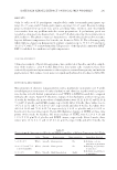
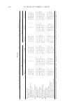
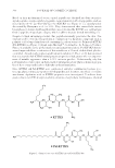
DATE PALM KERNEL EXTRACT ON FACIAL SKIN WRINKLES 279 containing 5% DPKE was prepared by the method described by Meer et al. (3). In brief, the cream was prepared as an oil-in-water disperse system by heating separately the aque- ous phase and the oil phase containing ABIL® EM90 as a nonionic emulsifi er (Cetyl PEG/ PPG-10/1 Dimethicone, Evonic Industries, Essen, Germany). The methanolic extract of date palm kernels was added in the aqueous phase, and the aqueous phase was added drop by drop to the oil phase at 75°C ± 1.0°C with continued stirring at 2,000 rpm. The emulsion was cooled at room temperature, and the speed of mixer was reduced gradually until the emulsion formed semisolid cream. The same method was used to formulate the base (placebo) cream without adding the DPKE. The composition of the two formula- tions is presented in Table I. The sta bility tests and the physicochemical characteristics of the test and placebo creams were performed at different time points during 8 weeks at 5 ± 0.1°C, 20 ± 0.1°C, 40 ± 0.1°C, 40 ± 0.1°C with 75% relative humidity, and 50 ± 0.1°C (9). PATCH TEST Before beginning the study, a patch test was performed on the skin of 12 volunteers (mean age 46.4 years) to investigate any skin reaction. A 5-cm area was marked on both forearms of each subject, and small amounts of both preparations were applied separately on each forearm then, the region was covered with surgical dressing. Positive skin reac- tions were carefully evaluated after 1, 48, and 72–96 h. The reaction was assessed accord- ing to the International Contact Dermatitis Research Group standard (10). TREATMENT PROTOCO L The region of int erest was the cheek and the temple areas. Both the cream form contain- ing DPKE and the placebo cream were coded and assigned to the right or left side of each volunteer’s face according to a computer-generated randomization list. Participants were asked to apply DPKE and placebo formulations separately on each side of the face twice a day for 8 weeks. Follow-up visits took place every 1 week throughout the study. CLINICAL ASSESSME NT Clinical assessme nt of facial skin was conducted as previously described by using three- point rating scales at baseline and at the end of the study (11). Skin roughness score: 1 = mild, 2 = moderate, and 3 = severe skin texture homogeneity score: 1 = slightly inhomo- geneous, 2 = uneven, and 3 = very uneven skin melanin pigmentation score: 1 = mild, Table I Cream Compositions Phase Placebo cream Cream with DPKE Oil phase Liquid paraffi n (17%) ABIL® EM90 (2.5%) Liquid paraffi n (17%) ABIL® EM90 (2.5%) Aqueous phase Distilled water (q.s 100%) DPKE (5.0%) Distilled water (q.s 100%)

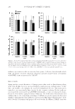
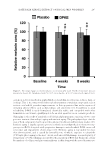
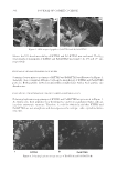
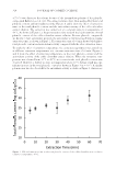
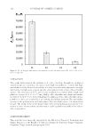
JOURNAL OF COSMETIC SCIENCE 280 2 = moderate, and 3 = severe skin redness score: 1 = mild, 2 = moderate, and 3 = severe. Assessment was performed to study subjects by two independent dermatologists, and repeat assessments were performed to judge the inter- and intrarater reliability, respectively. BIOPHYSICAL MEASU REMENTS Skin hydration, b arrier function, elasticity, pigmentation, and erythema were measured by a multiprobe adapter system (MPA system, Courage and Khazaka electronic GmbH, Koln, Germany). Skin surface hydration was assessed by a Corneometer probe (CM825) (12). The transepidermal water loss (TEWL) was assessed by a Tewameter probe (TM 300), which measures the density gradient of water evaporation from the skin. A micro- processor analyses the values and expresses the evaporation rate in g/m2/h (13). A Cutom- eter probe (MPA 850) measures skin elasticity based on the suction method. The settings of measurements were negative pressure 500 Mbar and suction for 3 s followed by 5 s of release. Both skin fi rmness and elasticity were measured (14). Skin pigmentation and erythema were determined with a Mexameter probe (MX18). Melanin measurement was calculated from the intensity of the absorbed and refl ected light at 660 and 880 nm, respec- tively, whereas erythema was measured at 568 and 660 nm (15). All probes were calibrated daily using standard references, and measurements were taken at baseline, after 4 weeks, and after 8 weeks. Values are displayed as arbitrary units (AU), and each value represents the mean of three measurements obtained from a participant’s same area of skin. SKIN SURFACE DIGI TAL PROFILOMETRY Skin surface profi lometry was performed by an Antera 3D® multispectral analyzer (Miravex Limited) at baseline and after 4 and 8 weeks of treatment. An Antera 3D® cam- era uses multidirectional illumination light to acquire multiple images and reconstructs a three dimensional image of the facial skin by the aid of computer software (8). After a period of 20 min acclimatization in controlled ambient conditions (24°C ± 1°C with 50 ± 10% relative humidity), the camera was placed directly on the external periorbital region over the crow’s feet lines of all subjects and optical scanning images were acquired. After the acquisition of images, several parameters related to skin topography were measured, including wrinkle depth, indentation index of fi ne lines (marginal size less than 1.5 mm), folds (marginal size less than 2.5 mm), and wrinkles (marginal size less than 5.0 mm). In addition, texture roughness, skin pigmentation (melanin), and redness (hemoglobin) were also assessed. The identical area was digitally identifi ed at each session, and values are presented as the result of triplicate measurements from the same area. STATISTICAL ANALYSIS Th e statistical analysi s was performed using SPSS version 15.0 (SPSS Inc., Chicago, IL) Data were represented as the mean ± SD. Multiple comparisons between means were performed as appropriate by using the paired t-test or the one-way analysis of variance (ANOVA) followed by Tukey post hoc test. In all the tests, a p-value of less than 0.05 was considered signifi cant.
Purchased for the exclusive use of nofirst nolast (unknown)
From: SCC Media Library & Resource Center (library.scconline.org)