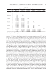
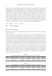
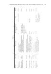
60 JOURNAL OF COSMETIC SCIENCE room temperature. A 0.3% caffeine gel (the amount of caffeine in tea extracts) and a gel- base were also prepared as described above. The contents of the gels were given in Table I. PHYTOCHEMICAL ANALYSIS Gels were weighed as 1 g and diluted to 10 mL in a beaker with distilled water. All solutions were filtered through a 45 µm filter before high-performance liquid chromatography (HPLC) analysis. The analytical procedure was adapted from the literature (15) with slight changes. The system was Perkin Elmer Series 200 and the column was C18 reversed-phase 5 μm (250 × 4.6 mm). System conditions were as follows: Mobile Phase A: 0.15% hydrochloric acid in water (v/v) Mobile Phase B: 0.15% hydrochloric acid in acetonitrile/water (v/v) Flow, 1 mL/min Injection Volume, 10 μL Column Temperature, 25°C. Detection was accomplished with a diode array detector and chromatograms were recorded at 280 nm. TOTAL PHENOLIC CONTENT ANALYSIS The total phenolic content of the extracts was determined using the Folin-Ciocalteu reagent (Sigma Aldrich, USA). The reagent was diluted at a volume ratio of 1:3 with 96% EtOH before use. Trolox (Sigma Aldrich, USA) was used as the standard. For this analysis, a calibration curve was made with 7 different concentrations of standard Trolox solution between 0.05–2.0 mM. The results were calculated using the regression equation of the obtained curve and defined as mg of Trolox equivalent, 1 g of tea gel was diluted to 10 mL with distilled water. 1 mL of this solution was mixed with 1 mL diluted Folin-Ciocalteu reagent, and 2 mL 35% sodium carbonate was added to the latter solution which was later diluted to 6 mL with distilled water. The final solution was incubated for 30 min at room temperature and the absorbance was measured at 700 nm. ANTIOXIDANT ACTIVITY ANALYSIS The cupric ion-reducing antioxidant capacity of samples was determined using an assay previously described (16). Briefly, 1 g of gel sample was diluted to 10 mL with distilled water. Subsequently 0.2 mL of 10 mM CuCl 2 ,0.2 mL of 7.5 mM neocuproine, and 0.2 mL of 1 M ammonium acetate were added into a test tube. After vortex mixing, a 100 µL sample, and 120 µL ultrapure water was added, and the absorbance at 450 nm was read Table I The Contents of the Gels g/100g Black tea gel Green tea gel Caffeine gel Vehicle gel Black tea extract 3 Green tea extract 3 Caffeine 0.3 Carbomer 1 1 1 1 Sodium hydroxide qs qs qs qs Benzyl alcohol 1 1 1 1 Pure water qs qs qs qs qs: quantum sufficit.

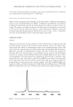
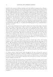
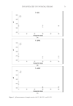
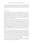
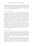
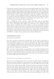
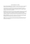
61 PRELIMINARY SCREENING STUDY WITH TEA FORMULATIONS 30 min later. Trolox equivalent antioxidant capacity was calculated based on a calibration curve obtained by the serial dilution of 1 mM Trolox. FREE RADICAL SCAVENGING ACTIVITY ANALYSIS The free radical scavenging activity of samples was measured with 1,1-diphenyl-2 picrylhydrazil using an assay described formerly (17). Briefly, 1,1-diphenyl-2 picrylhydrazil was dissolved in ethanol (4 mg/100 mL) and 100 μL of this solution was added to an equal volume of a sample. The mixture was shaken vigorously and the decrease in absorbance was measured at 515 nm after 30 min. Water was used as a control. The percent inhibition activity was calculated using the following equation: Inhibition activity (%)=[(A 0 – A 1 )/A 0 × 100], where A 0 and A 1 are the absorbances of the control and sample, respectively. CLINICAL STUDY SUBJECTS The group consisted of 21 caucasian subjects, 15 females and 6 males, 20–48 years old, with no known disease enrolled in the trial with written informed consent. The subjects were chosen under the control of a dermatologist based on the inclusion/exclusion criteria: The subjects were not using any topical or systemic medications, they had all healthy dermatologic appearance, they had no photosensitivity history, and none of them were pregnant or lactating. All of the trial procedures were performed in line with the ethical principles laid down for medical research (Helsinki Declaration of World Medical Association, 1964, and amendments). The study protocol was approved by the local ethics committee. After grading the skin types, the minimal erythema dose (MED) of each subject was determined on the upper back skin with the artificial narrow band UVB source (Gigatest UVB-311, Germany) and its emission spectrum is shown in Figure 1 (18). This handheld MED tester has five test fields, each with a diameter of 15 mM which emits different doses of UVB. The MED was determined as the lowest UVB dose that caused just perceptible erythema at 24 h. Figure 1. The emission spectrum of the lamp (UVB) (18).
Purchased for the exclusive use of nofirst nolast (unknown)
From: SCC Media Library & Resource Center (library.scconline.org)