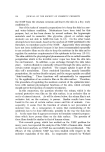
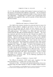
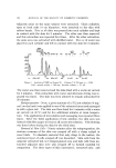
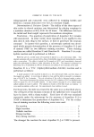
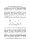
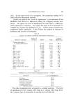
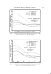
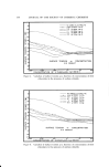
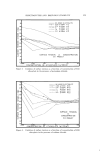
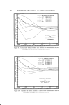
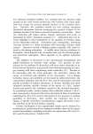
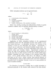
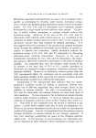
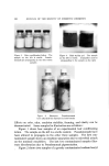
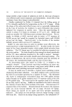
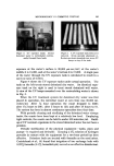
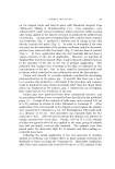
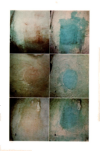
DERMAL IRRITATION 201 on the clipped backs and held in place with Blenderm Surgical Tape (Minnesota Mining & Manufacturing Co.). Test substances were utilized which, under normal eonditions• either caused no visible reaction after being applied to the skin for 24 hours or produced the mildest type of erythema. An injection of Sulphan Blue into a rabbit whose response was borderline (Fig. 1) produced a definite area of change in the skin directly beneath the patch (Fig. 2) In order to determine if the time necessary for the formation of the primary erythema could be decreased• patches were removed after four hours (Fig. 3) and one hour of contact (Fig. 5). At these application times the test materials did not show a visual erythema at the site of application. However, an injection of Sulphan Blue into these animals (Figs. 4 and 6) showed a definite increase in the intensity of the dye at the site of product application. This indicated that changes were occurring in the skin, as evidenced by the concentration of the dye this• in turn, could be correlated with mild erythema which could not be seen without the aid of the injected dye. Vinson and Borselli (5) recently outlined a method for developing photosensitization in the guinea pig. It was felt that there was a need to re-examine this method to, i• determine if the procedure and response could be duplicated using strains of animals other than the closed strain which was employed by the authors• and• ii, whether the use of Sulphan Blue could increase the sensitivity of the test. Guinea pigs were purchased from three commercial breeders, and the procedures followed were essentially those described in the published paper (5). Groups of five animals of both sexes each received fl.05 ml of a 2% solution in alcohol of either Bithionol or Temasept II. After application of the test material to the clipped cervical region, the animals were exposed for 15 minutes to a No. 27,5 Westinghouse Ultraviolet bulb at a distance of 46 cm. This procedure was repeated daily for five consecutive days. After the last treatment the animals were allowed to remain untreated for seven days. Finally• 0.05 ml of a 0.1% solution of either test agent in olive oil was applied to the same group of animals that had previously received this material. The animals were then placed under the ultraviolet light for 15 minutes and skin readings re- corded 24 hours later. Following the initial application of the test materials in absolute alcohol no erythema was evident either in those animals receiving the Bithionol or those receiving the Temasept II. Ultraviolet irradiation, after these same animals had received 0.05 ml of a 0.1% solution of the

2O2 JOURNAL OF THE SOCIETY OF COSMETIC CHEMISTS test agent in olive oil, did cause mild erythema in those animals which had been treated with Temasept II. After the skin responses had been recorded, each animal received an intraperitoneal injection of l ml of a 6% solution of Sulphan Blue. Within ten minutes after the injection the dye had accumulated at the sites of application of the Bithionol but not at the sites of application of the Temasept II. Since the response reported was obtained in animals from three different sources it may be concluded that the method of Vinson and Borselli can be utilized to screen products which may induce photosensitivity. SUMMARY Sulphan Blue should be considered as a useful adjunct in evaluating those materials which produce a minimal degree of vasodilation which is normally not readily discernible in rabbit patch tests or the guinea pig photosensitivity tests. (Received August 22, 1966) REFERENCES (1) Taintcr, M. L., Throndson, A. H., and Lehman, A. J., Local irritation from sodium bisulfite as preservative in epinephrine solutions, Proc. Soc. Exp. Biol. Med., 36, 584 (1937). (2) Hoppe, J. O., Alexander, E. B., and Miller, L. C., Use of the Trypan Blue and rabbit cyc tests for irritation, J. Pharm. Sci., 39,147 (1950). (3) Finkclstcin, P., Laden, K., and Micchowski, W., Laboratory methods for evaluating skin irritancy, Toxicol. Appl. Pharmacol., 7, Supplement 2, 74 (1965). (4) Brown, V. K., and Clark, R. A., Sulphan Blue as an aid to the laboratory assessment of primary skin irritants, J. Invest. D ermatol., 45,173 (1965). (5) Vinson, L. J., and Borselli, V. F., A guinea pig assay of the photosensitizing potential of topical germicides, J. Soc. Cosmetic Chemists, 17,123 (1966). Figure 1. Borderline reaction. Figure 2. Borderline reaction following Sulphan Blue injection Figure 3. Erythema after four hours Figure 4. Erythema after four hours following Sulphan Blue injection Figure 5. Erythema after one hour Figure 6. Erythenla after one hour following Sulphan Blue injection
Purchased for the exclusive use of nofirst nolast (unknown)
From: SCC Media Library & Resource Center (library.scconline.org)