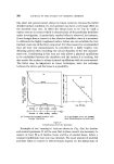
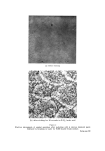
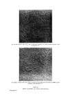
418 JOURNAL OF THE SOCIETY OF COSMETIC CHEMISTS about, but one possible means of affecting this would be by the action of these high-level dendrocytic cells. It is beyond reasonable doubt that the autolysis of the cell protoplasm prior to keratinization is due to hydrolytic enzymes released in the upper layers of the epidermis at the level of cells known as the granular layer. In skin diseases where there is no organised liberation of these enzymes, none of the cell contents is removed, and the whole of the cell, including its nucleus, becomes involved in the process of keratinization. Such a state of affairs exists in the skin disorder, psoriasis. Palmar and plantar keratinization occupy a position midway between the keratin produced in psoriasis and that, for example, produced on our backs. Here much of the cell contents, except the nucleus, escape auto- lysis and consequently the keratin layer is more solid and stable than that formed on the back. It is important to mention that although the keratin layer is different this does. not necessarily imply that the actual keratin molecules forming the horny layer differ from each other. As we have detected three distinct enzyme systems in melanocytes other than their dopa oxidase activity, it would perhaps seem reasonable that this other hydrolytic activity might influence the mode of keratiniza- tion of the epidermal cells. The ATPase activity is probably related to motility of the cells. It is most unlikely that it is the type of ATPase associated with ion transfer through cell membranes, and is probably the type associated with contrac- tile protein. This would suggest that the dendritic cells move actively up within the epidermis and are not passively carried upwards by the general movement of the epidermal cells. Acid phosphatase and sulphatase are both lysosomal types of hydro- lytic enzymes, and both would be capable of causing cellular hydrolysis if they were transferred into the epidermal cell in a similar manner to that in which the melanin granules are transferred into epidermal cells of the basal region. It is also possible that any effects of these high-level den- drites could be due to an induction or activation of hydrolytic enzymes rather than to an actual transfer of such an enzyme into epidermal cells. Although the precise mechanism cannot be directly demonstrated, the circumstantial evidence points strongly towards the involvement of the high-level dendrocytes. Thus in plantar skin where there is relatively little hydrolysis, it is extremely difficult to demonstrate ATPase active dendritic cells, whereas in normal epidermis where there is a much greater hydrolysis of the epidermal cells they are readily detectable in considerable numbers.

THE MELANOCYTE SYSTEM AND KERATINIZATION 419 In psoriasis, which is associated with the absence of organised hydro-. lytic enzyme activity, ATPase cells are virtually absent in the lower layers of the epidermis and those in the upper part of the epidermis are abnormal in that they show weak ATPase activity and are without dendrites. Acid phosphatase and sulphatase have not been demonstrated within dendritic cells although they are profusely scattered throughout the psoriatic epi- dermis. Comparative studies on lower mammals also support the hypothesis that the dendritic cells have an influence on the type of keratin produced by the epidermal cells. (Received: 8th March 1966) REFERENCES (1) Masson, P. in The biology ofrnelanornas 4 15 (1948) (N.Y. Acad. Set.). {2) Jarrett, R. and Riley, P. A. Brit. y. Dermatol. In the press. (3) Fitzpatrick, T. B., Brunet, P. and Kukita, A. in Ellis, R. A. and Montagna, W. (eds.) The biology of hair growth (1958) (Academic Press, New York). (4) Billingham, R. E. and Medawar, P. B. _Phil. Trans. Roy. Soc. London Ser. B. f•l? 151 (1953). (5) Jarreft, A. and Riley, P. A. Brit. J. Dermatol. ?õ 79 (1963). (6) Jarrett, A. and Spearman, R. I. C. Histochemistry of the skin: _Psoriasis. (1964) (English Universities Press, London). DISCUSSION DR. L. GOLBER6: A substantial proportion of the population are taking phena- thiazines in one form or another, or are exposed to things like chloroquin and it is now suggested that all new drugs should be screened for their ability to combine with melanin as a protective measure against the possibility of damage to the retina. In the skin we have another situation where there is melanin and the possibility of combination with it we know that molecules of this sort also tend to be selectively accumulated in lysosomes and there would be a tendency for such substances to accumulate in the skin. On numerous occasions there have been clinical descriptions of skin staining developing, say, from the administration of phenathiazine, but to what extent are there minimal degrees of change in skin pigmentation which are not so striking as to draw themselves to the attention of the physician, but yet do affect the shade of the skin ? THE LECTURER: Lysosomes are at the present time one of our interests and much of this work on sulphatases and the other acid hydrolases are in point of fact directed to the lysosomal activity of the skin. There is good reason for believing that the skin hydrolases are actually of lysosomal origin. Although the electromicroscopists have for years stoutly denied that they have been able to detect lysosomes in epidermal cells, they are beginning to change their minds. They have now definitely stated that these bodies are present in high level melanocytes, and more recently a paper has been published in which they have admitted seeing them in the epidermis from cases of atopic eczema, but they still deny having seen them in normal skin. However, I feel sure they must be there, at least in the sense that the lysosome is only a concept of containing active hydrolyric enzymes within protective envelopes. The fact that their pictures do not show the usual correct anatomical size or shape of envelope does not
Purchased for the exclusive use of nofirst nolast (unknown)
From: SCC Media Library & Resource Center (library.scconline.org)