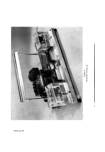
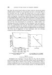
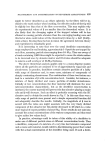
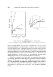
416 JOURNAL OF THE SOCIETY OF COSMETIC CHEMISTS A photographic technique was also developed in which a uniform radiation of the skin with uv rays was attained by a battery of four uv lamps (Osram 125 MEW/V) having a total output of about 14 watts at a wavelength of about 3,650 A. The area of skin under investigation was photographed through an OX1 1 mm filter at a standard distance and exposure, and the emulsion processed to a given gamma. Densitometry readings of the negative, compared with a simultaneously photographed control, were made with a radiological densitometer. This method gave comparable results with those obtained by reflectance photospectometry. THE NATURE OF SKIN PIGMENT Melanin, as already mentioned, is formed by specific epidermal cells known as melanocytes. The only criterion of a melanocyte is that it produces melanin, and at present the only satisfactory method for its histo- logical demonstration is by incubating epidermis in dihydroxyphenylalanine (dopa). It is remarkable that this compound should be more readily acted upon by the enzymes of the melanocyte than the postulated natural substrate, tyrosine, which is thought to be the true precursor of melanin. The reason given for this anomaly is that in vitro the melanocyte is unable to accomplish the first stage of transforming tyrosine into "dopa" and if, therefore, "dopa" is presented to the cell the rest of the metabolic pathway towards melanin formation proceeds to completion. As mentioned above, the precursor of melanin is thought to be tyro- sine, and by a series of reactions, some enzymatic oxidations by the enzyme tyrosinase and some autoreductions, this amino acid is transformed into the brown-black pigment, melanin. In the final product this is closely associated with protein to form melanin granules, which are then passed into adjacent epidermal cells. Another type of melanin has also been described, known as pheomelanin (3). This is the yellow-coloured pigment which is thought to be responsible for the yellow skin colour of the Oriental races and also for the yellow colour in the hair of "agouti" varieties of several animal species. Some authorities consider that the production of pheomelanin involves similar metabolic pathways to those of melanin synthesis and that possibly it is derived from the same precursor, tyrosine. A difference between these two melanins is shown by their solubilities in sodium hydroxide. It has also been suggested that pheomelanin represents a further oxidation product of melanin.

THE MELANOCYTE SYSTEM AND KERATINIZATION 417 OTHER ASPECTS OF MELANOCYTE FUNCTION The definition of a melanocyte is that it is a cell that forms melanin and is able to oxidize dihydroxyphenylalanine to a black pigment. It has also been shown that dendritic cells could be detected at higher levels in the epidermis than the basal layer. These cells situated in the upper regions of the epidermis were, however, dopa negative. Therefore other methods had to be used for their demonstration and the gold impregnation tech- niques were probably the most reliable for their detection. Because these so-called high-level melanocytes were dopa-negative it was considered that they were melanogenically effete and had no further function. They were carried passively upwards with the general movement of the epidermal cells, and were finally exfoliated with the keratin layer. Recently we have used various enzyme techniques on human and animal skins and were surprised to find that although these high level cells were inactive in respect to dihydroxyphenylalanine, nevertheless they were very active in respect to other substrates. The first enzyme to be detected was adenosine triphosphatase (5). Later we obtained evidence that they also exhibit acid phosphatase and sulphatase activity. It would, therefore, appear that while they no longer produce pigment they almost certainly perform other functions at higher levels in the epidermis. During the process of keratinization the epidermal cells, or keratino- cytes as they are sometimes called, become transformed into a stable, chemically-resistant fibrous protein known as keratin. This unreactive material, together with its contained lipids, acts as the first barrier against physical and chemical insults to the skin in particular, and to the body as a whole. The process of keratin formation appears to be a two-fold process. One is cellular cytolysis in which the nucleus and other components of the cell are removed before the occurrence of the second stage of molec- ular keratin polymerization. Thus, the physical nature of the final keratin layer can be modified to the particular site rcquirements of the animal depending on the relative amounts of the celhflar •naterial removed before keratinization (6). It is quite obvious that the horny layer on our palms and soles is differ- ent from that of other regions, but it can be shown that more subtle differences also exist between other skin sites. It is difficult to explain the mechanism whereby these regional differences in keratinization are brought
Purchased for the exclusive use of nofirst nolast (unknown)
From: SCC Media Library & Resource Center (library.scconline.org)