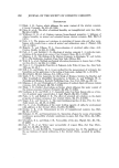
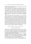
9,48 JOURNAL OF THE SOCIETY OF COSMETIC CHEMISTS .5 20 15 lO 0 200 400 600 800 l OO0 1200 1400 1600 1800 2000 Time {sec.) Figure 5. Water absorption of stratum corneum at several RH's as a function of time - ß- ß - ß-- 93% RH unextracted * * * 93% RH extracted -I-ffi-ffi- 60% RH unextracted -}--•-•-30% RH extracted Sample Table II Equilibrium Moisture Content of Haman Stratum Corneum % Water Absorbed by Dry Stratum Cometun a % RH Unextracted Extracted A A B B C C C C C C C 31 5.4 o (4.9-6.0) 76 11.4 ø (10.8-12.1) 31 9.36 (8.5-10.3) 76 13.1 a (12.0-14.2) 5 2.5 15 4.0 31 4.0 35 6.5 50 8.66 (7.9-9.3) 75 14.5 • (13.8-15.1) 90 21.9' (21.'8-22.1) "Figures in parentheses are ranges. øAverage of 5 determinations. CAverage of 3 determinations. SAverage of 4 determinations. *Average of 2 determinations. 5.3 ø (3.9-6.2) 13.3 ø (12.5-14.1) 6.5 a (5.8-7.3) 14.1 a (12.8-15.0) 2.5 15.0 5.1 7.0 9.9 ø (9.0-11.0) 15.8' (14.2-17.4) 22.5 ø (20.4-24.5)

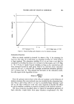
WATER LOSS OF STRATUM CORNEUM 249 Water Vapor Transmission The results of in vitro transepidermal water losses through untreated stratum corneum at 21---1.5øC against a dry atmosphere (CaC12) show wide variations depending on the stratum corneum specimen. When watet vapor is in contact with the stratum corneum inside the cell, the average rate is about 0.25 mg cm-"' hr-L When water contacts the stratum corneum, the average transepidermal water loss rises to about 0.30 mg cm -2 hr -•. These values are fairly close to each other and are in accord with normally accepted values of transepidermal moisture loss obtained in vivo (19). Extraction of stratum corneum, first with pyridine and then with water, in- creases the rate of transepidermal moisture loss from about 0.2 to as much as 1.6 mg cm -2 hr '•. This is a clear indication that the extractables (primar- ily lipids) present in the stratum corneum contribute significantly to slowing down the moisture loss from dermal and epidermal tissue by evaporation. On theoretical grounds and intuitively one would expect that the trans- epidermal water loss from a cell of the type described here depends on the RH to which the cell is exposed, i.e., the driving force for diffusion is the chemical potential gradient. That this is the case has been shown here be- cause the plot of water vapor transmission rate vs. RH (Fig. 6) is linear with a slope of -1.95 x 10³ mg cm-"' hr-•/% RH. These results are different from those reported by Grice et al. (2), who indicated that a plot of water vapor transmission vs. RH yields a curve showing a maximum between 25 and 50% RH (Fig. 6). It was at first thought that this difference might be due to the fact that an unstirred layer in the system used here was responsible for the discrepancy. However, when the results were repeated under a stream of air (1200 ml/min) aimed at the exposed surface of the stratum corneum, the results were essentially unchanged. It appears, therefore, that the results of the in vitro experimentation conducted here conform to the in vivo data reported by Goodman and Wolf (15), whose results yield linear plots al- though the slope of their line has a value of -4.5 x 10 a mg cm -• hr-•/% RH. The effect of temperature on the permeability of stratum corneum or any membrane follows the Arrhenius equation (oe0). Thus, a plot of the 1 Ep logarithm of permeability vs.•, is linear and has a slope of 2.303R' where E• is the activation energy of permeation. The experimentally measured flux, J, is related to permeability (P) via eq 3, (3) where AC = the difference in the concentration of the water on the internal side of the membrane and the external side (moles 1-•).
Purchased for the exclusive use of nofirst nolast (unknown)
From: SCC Media Library & Resource Center (library.scconline.org)