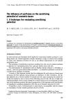
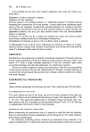
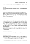
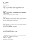
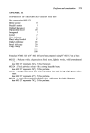
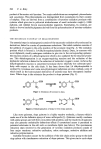
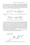
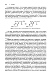
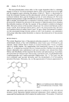
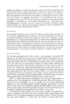
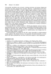
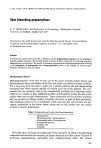
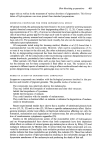
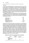
The mechanism of skb, pigment production 397 (Fig. 3). This compound is probably identical to the leucodopachrome of Raper (2). Under appropriate conditions there is a spontaneous rearrangement of cyclo-dopa with the loss of carbon dioxide and two hydrogen atoms to give 5,6 dihydroxyindole (Fig. 4). There is some evidence to suggest that this dehydrogenation is achieved by a redox exchange with a quinone intermediate. In the absence of this step alternative additive reactions may give rise to oligomers containing cyclo-dopa (3). o ••-• co OH 0 HOo•COOH H H Figure 3. Cyclization of dopa quinone to give the indolene product cyclo-dopa (leucodo- pachrome). COOH -C02 HO H -2H H Figure 4. Rearrangement of cyclo-dopa to give 5,6-dihydroxyindole. In addition to catalysing the conversion of cyclo-dopa the quinone intermediates, such as dopa quinone and indole 5,6 quinone, may take part in the oxidation of compounds which cannot be directly oxidized by tyrosinase by virtue of their structure or their location in relation to the enzyme. Some of these redox reactions may be extremely deleterious to the pigment-producing cells (for discussion see ref. 1). In man at least one mechanism is known to exist which traps the reactive quinones and prevents them from initiating cellular damage. The trapping reaction consists of the formation of a reduced C-5 adduct with glutathione. The glutamic acid and glycine residues are cleaved by peptidases giving rise to 5-S-cysteinyl dopa, whicl• can be detected in melanocytes and in the serum, and is excreted in the urine (4). The melanin group at Naples (5) have shown that cysteinyl dopa can also be formed by a direct interaction between dopa quinone and cysteine (Fig. 5). O• COOH 0 •"•"•--• N H 2 HOOC• s HOOC• SH Figure 5. Condensation of dopa quinone with cysteine to form 5-S-cysteinyl dopa.

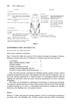
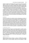
398 P. A. Riley Two isomers are formed in vitro: 2-cysteinyl dopa and 5-cysteinyl dopa, the latter in proportionately much larger amounts. If the product is oxidized to cysteinyl quinone spontaneous cyclization takes place with reduction to dihydrobenzothiazine (Fig. 6). This reaction is linked to the oxidation of a diphenol such as cysteinyl-dopa so that the generation of dihydrobenzothiazine, once initiated, can proceed non-enzymatically. This may explain the apparently paradoxical findings of Prota's group (6) that 3-x•C - cysteine incorporation by pigment cells is related to tyrosinase activity whereas Gesch- wind, Huseby and Nishioka (7) showed that MSH administration to A ¾ mice switched melanocytes from pheomelanin to eumelanin production. o•CO OH Ho•CO OH -,. Figure 6. Reduction of 5-S-cysteinylquinone to form dihydrobenzothiazine. It has been shown that the pheomelanins are composed of more or less complex polymers of dihydrobenzothiazine. A number of trichochromes which are composed of dimers of dihydrobenzothiazine have been identified and degradation studies of gallo- pheomelanin obtained from New Hampshire hen feathers have shown it to be an irregular and complex polymer perhaps containing benzothiazole and tetrahydroisoquinoline ring systems. A similar lack of simplicity is evident in the composition of eumelanins. Polymeriza- tion of the oxidation products of tyrosine takes place by a series of reductive additions. A random assortment of oxidized intermediates is incorporated into the polymer which therefore lacks any specific structure. The most regular structure is one produced by polymerization between C4 and C7 atoms of adjacent indole quinone rings. X-ray diffraction studies indicate that stacking of the rings occurs forming a lattice arrangement and this constraint may be responsible for introducing some regularity into the polymer by restricting access to sterically affine structures. Two major differences between eumelanins and pheomelanins emerge from a knowledge of their biosynthesis. Pheo- melanins, because they are polymers of less conjugated precursor molecules, show a much more restricted spectral absorbance than eumelanins and thus give the structures which they pigment a reddish or yellowish appearance in contrast to the dark brown or black of the highly conjugated eumelanins. Secondly, because the cyclization of cysteinyl-quinone replaces one of the hydroxyl groups on the phenol ring, the pheomelanin polymer cannot contain reactive quinones and this prevents the formation of cross-linkages with protein and leads to the characteristic lack of organization within pheomelanin pigment granules. In the case of eumelanins the protein tanning effect of quinone constituents of the pigment is very marked and seems to be of importance in the hardening of insect cuticles and sclerotization reactions in other invertebrates (1). In mammalian melanosomes the reaction of eumelanin quinones with protein probably accounts both of the melanin deposition on the matrix and the inhibition of tyrosinase in the fully melanized granule. The highly conjugated structure of eumelanins permits electron movements and electron exchanges with neighbouring molecules take place readily, i.e. melanins are chemically highly reactive. Commoner, Townsend and Pake (8) first demonstrated the paramagnetic
Purchased for the exclusive use of nofirst nolast (unknown)
From: SCC Media Library & Resource Center (library.scconline.org)