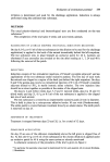
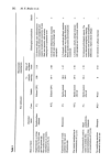
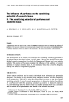
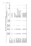
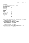
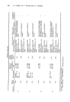
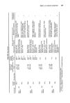
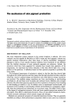
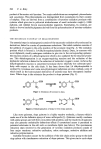
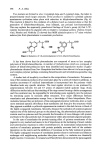
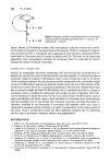
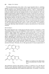
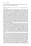
The mechanism of skin pigment production 399 properties of melanins and subsequently Mason's group (9) showed that the free radical property of melanin is due to semiquinones, stabilized by resonance in the highly con- jugated polymer and steric restrictions on internal radical annihilation reactions. Mole- cular orbital calculations by Pullman and Pullman (10), based on an assumed structure for dopa-melanin, showed that several redox states are possible and predicted electron acceptor properties which have been confirmed by several studies (11, 12, 13, 14). This reactivity of melanins may be the reason which necessitates its compartmentation in membrane-bounded melanosomes in the melanocytes (15). When transferred to the cytoplasm of keratocytes, melanin granules probably act as initiators of cytoplasmic damage in cells exposed to radiation which is absorbed by the pigment and generates free radicals (9, 16). The evidence for this proposal is discussed elsewhere (1, 17, 18) but it is probable that the selective advantage to hairless mammals is the destruction of cells which have received a radiation dosage sufficient to cause deleterious mutations. Clearly, from what has been said about their structure, the pheomelanins are much less effective in this respect and this is borne out by the statistics on the susceptibility of various ethnic groups to skin cancer. TYROSINASE The enzyme responsible for the oxidations involved in melanogenesis is tyrosinase. At least two forms (a and [•) of the enzyme are recognized and minor differences in activity exist but the general properties are broadly identical. They are widespread in nature, occurring in both eukaryotic and prokaryotic organisms. In vertebrates the enzyme is synthesized only in specialized cells (melanocytes) and is active only in specialized cyto- plasmic organelles (melanosomes). In contrast to many other enzymes whose structures have been determined and for which the catalytic mechanisms are well understood, tyro- sinase is still very poorly comprehended and no clear reaction mechanism has emerged. It is known that tyrosinases contain copper and that they bind oxygen. There are two main classes of oxidation catalysed by tyrosinases: the dehydrogenation of diphenols (catecholase activity) and the ortho-hydroxylation of monophenols (cresolase activity). CATECHOLASE ACTIVITY The enzyme isolated from Neurospora crassa has been studied by a number of groups. The enzyme has a molecular weight of about 33 000 daltons and contains one atom of copper per mole of protein (19). The kinetic studies of Gutteridge and Robb (20) in- dicate a single binding site for diphenolic substrates. Electron spin resonance studies show that only 4•o of the copper is in the cupric form and spectral absorbance data indicate that 35•o is complexed with monomolecular oxygen leaving about 60•o of the enzyme free to combine with the substrate. Binding to substrate and oxygen is thought to take place in a random sequence (Fig. 7). The Neurospora enzyme appears to be incapable of oxidizing monophenols. CRESOLASE ACTIVITY Mushroom (Agoricus bisporus) tyrosinase is a tetramer of 130 000 daltons molecular weight and contains four atoms of copper. On the basis of the reaction with hydrogen peroxide, Jolley et al. (21) have proposed a dimeric active site containing two copper

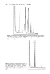
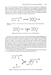
400 P. A. Riley •,,...• ' •,•..,•I 0 + Q + HzO I:'DO E + Q + H•O Figure 7. Schematic outline of catecholase activity of Neurospora tyrosinase (after Gutteridge and Robb (20). E = enzyme diphenol Q = quinone. atoms. Mason (22) furnished evidence that monophenol oxidation involves the transfer of two electrons to generate the cupric form of the enzyme. If this is confirmed it suggests that cresolase activity is consequent upon aggregation of monomeric catecholases of the type found in Neurospora and Streptomyces glaucescens (23). This leads to the interesting speculation that monophenol oxidation by tyrosinase could be controlled by factors limiting the extent of subunit interaction. MAMMALIAN TYROSINASE Studies on mammalian tyrosinase employing SDS poly-acrylamide electrophoresis by Burnet's group (24) have shown that the smallest sub-unit capable of oxidizing dopa has a molecular weight of roughly 65 000 daltons -which would correspond in size to the dimer of the Neurost•ora enzyme. In some instances another component with a molecular weight of about 120 000 daltons can also be detected which is about the size of the tetramet of the mushroom enzyme. However, a principal component in the material obtained from mice has a molecular weight of about 80 000 daltons and it is probable that this is a form of tyrosinas½ which is modified by interaction with another protein component. The struc- ture of tyrosinase is under the control of the C (or colour) locus. Modifying loci, such as pink-eyed dilution and dilute, seem to have the effect of converting more of the enzyme into the modified form. It may be that the coloration of animals carrying these genes is, therefore, modified by an interaction of tyrosinas½ with peptides which may cause a reduction in cr½solase activity, possibly as a result of a separation of the copper atoms in the dimer. Such a modification would not affect diphcnol oxidation but, by inhibiting the first step in tyrosinas½ oxidation, reduce overt pigmentation. REFERENCES 1 Riley, P. A. The mechanisms of melanogenesis. Symt•. Zool. Soc. Lond. 39 77 (1977). 2 Raper, H. S. The aerobic oxidases. Physiol. Revs. 8 245 (1928). 3 Gruhn, W. B., Pomeroy, J. S. and Maurer, L. M. An oligomeric hydroxyphenylalanine in malignant melanoma: a new type of melanogen. Biochem Biophys. res. Comm. 61 704 (1974). 4 Rorsman, H. The melanocyte illuminated. Trans. St John's Hosp. Derre. $oc. 60 135 (1974). 5 Prota, G. and Thomson, R. H. Melanin pigmentation in mammals. Endearour :t5 32 (1976). 6 Misuraca, G., Nicolaus, R. A., Prota, G. and Gliara, G. A cytochemical study of pheomelanin formation in feather papillae of New Hampshire chick embryos. ExI•erientia 25 920 (1969). 7 Geschwind, I., Huseby, R. A. and Nishioka, R. Recent Progress in Hormone Research Vol. 28, pp. 91-130, Academic Press, N.Y. (1972). 8 Commoner, B., Townsend, J. and Pake, G. E. Free radicals in biological materials. Nature 174 689 (1954).
Purchased for the exclusive use of nofirst nolast (unknown)
From: SCC Media Library & Resource Center (library.scconline.org)