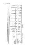
J. $oc. Cosmet. Chern. 29 25-29 (1978) Towards objectivity in the assessment of eye irritation R. HEYWOOD and R. W. JAMES Huntingdon Research Centre, Huntingdon, PE18 6ES Synopsis The assessment of eye irritation is subjective. The eye test system is subject to such wide variation that it will never be possible to make precise measurement of irritancy. Clinical appraisal, supported by measurement of comeal thickness and intra-ocular pressure, is probably the best that can be achieved. The use of local anaesthetics should be considered when carrying out eye irritation tests in the rabbit. INTRODUCTION The assessment of eye irritatation is basically subjective and it is not surprising that the literature abounds in results showing different inter- and intra-laboratory values. The need for objective assessment has been voiced by many investigators and some para- meters for the measurement of functional and pathological change have been proposed. The purpose of this paper is to review the feasibility of some of these methods. CLINICAL EXAMINATION The assessment of irritants is based on the well-established Draize test- Draize, Woodard and Calvery (1)- which has been modified over the years, although most laboratories follow the standard procedure laid down by the U.S.A. Code of Federal Regulations (2). Albino rabbits of either sex weighing between 2 and 3 kg are the usual test species. It must be pointed out that differences due to age, sex, management, strain, season and other environmental factors have received scant attention, although there is little doubt that the strain of rabbit is of considerable importance. The degree of damage induced is dependent on the concentration of compound and the intimacy and duration of contact with the conjunctiva and cornea. The concentration of compound used should be that which induces a minimal measurable response. The intimacy of contact can be controlled by applying a known volume and standardising the application procedure. The other factor that could be varied is the period of contact between the test material and the conjunctival sac. It has been shown by Davies, Kynoch and Liggett (3) that 10 sec is the maximum delay time, after which irrigation is not beneficial. This initial contact time is too critical to control and it is inappropriate to introduce a washout technique and so add another variable to what is already a compli- cated situation. Another factor against initiating washout techniques is the fact that the washing procedure is not always beneficial. Sebaugh et al. (4) showed that the washing procedure shortened the onset of opacities produced by weak acids, but with 1•o 0037-9832/78/0100-0025 $02.00 ¸ 1978 Society of Cosmetic Chemists of Great Britain 25

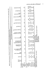
26 R. Heywood and R. W. James sodium hydroxide washing enhanced the initial eye irritation. With strong acids and alkalis, washing has no effect. The response of the conjunctiva, cornea and iris are graded on a 0-4 scale. It is at this point that observer bias comes in. The reactions are best scored by technical staff trained to a common standard. Bayard and Hehir (5), investigating reader-scoring variability, found that readers were consistently able to produce a positive or negative reading more than 90•o of the time. If clinicians, be they veterinary or medical, examine reactions, they are biased by their experience of clinical conditions in the eye and tend to score to lower grades than technical staff. Ballantyne et al. (6) suggested grading for lachrimation, blepharitis, chemosis, hyperaemia, sloughing, iritis, keratitis and for corneal vessels. It is common practice to manipulate the scores in order to express the total irritancy as a single number. Attractive as the idea may be to express a complicated biological situation as a single number, the practice must be viewed with caution. Ballantyne and Swanston (7) state that it is not possible to grade eye irritancy simply on the grounds of numerical values and that observed effects must be summarised verbally. There is considerable variability between individual animals, whether one is working with rabbits or monkeys. To reduce the number of animals would decrease the ability of the test to differentiate degrees of irritation, but increasing the group sizes would not increase precision. Comparison of the total Draize score and scores from the cornea only suggest that the conjunctiva makes the initial response to irritation and that the reaction persists for longer than it does in the cornea. The effect of this conjunctival response is not under- stood, but it could be considerable. The present trend in Draize testing is to reduce concentrations of the working solutions in the hope of being able to measure conjunctival response only. Despite our concern about the Draize test, Marzulli and Ruggles (8) reported that reliable and reproducible results could be obtained in different laboratories in distin- guishing an irritant from a nonirritant. They recommend a simple pass/fail criterion, monitoring four parameters. SLIT LAMP EXAMINATION In our experience, this examination, together with fluorescein staining, is the most sensitive indicator of corneal damage in the monkey. If fluorescein is instilled into the conjunctival sac of the rabbit, in nearly all cases staining patterns will result. Kikkawa (9) described intervals of 1-11 days between light and intense staining and correlated the pattern in both eyes. The stain pattern was attributed to physiological desquamation of the corneal epithelium. In our experience, this pattern of intense and light staining has not been established in the rabbit. The turnover rate of epithelial cells in the cornea is high and the vulnerability of the corneal epithelium to insult may reflect the different stages in the life-cycle of the cells. Nevertheless in monkeys this high turnover of cells is achieved without a break in the continuity of the epithelium. The fact that the corneal epithelium of the rabbit is not intact will generally increase the rate and amount of penetration, this being particularly true for water soluble and polar compounds. CORNEAL THICKNESS Corneal thickness can be readily measured following the technique defined by Mishima and Hedbys (10) using the Haag-Streit depth measuring attachments. Burton (11) must
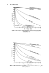
Purchased for the exclusive use of nofirst nolast (unknown)
From: SCC Media Library & Resource Center (library.scconline.org)