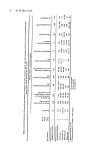
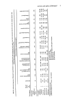
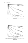
Objectivity in assessment of eye irritation 29 that the eye test system is subject to such wide variation that it will never be possible to make precise measurements of irritancy and that it will always be a subjective clinical appraisal, supported by measurements of corneal thickness and probably of intraocular pressure. Certain aspects of irritancy, such as the 'sting' phenomenon, which is probably of major commercial importance, cannot be measured in animal systems. REFERENCES 1 Draize, J. H., Woodard, G. and Calvery, H. O. Method for the study of irritation and toxicity of substances applied topically to the skin and mucous membranes. J. Pharmacol. Exp. Therap. 82 377-389 (1944). 2 Code of Federal Regulations. Tests for eye irritants. Title 16, Section 1500. 42 (1973). 3 Davies, R. E., Kynoch, S. R. and Liggett, M. P. Eye irritation tests - an assessment of the maximum delay time for remedial irrigation. J. Soc. Cosmet. Chern. 27 301-306 (1976). 4 Sebaugh, V. M., Osterberg, R. E., Hoheisel, C. A., Murphy, J. C. and Bierbower, G. W. A compara- tive study of rabbit ocular reaction to various exposure times to chemicals. Society of Toxicology JSth Annual Meeting Abs. 237 (1976). 5 Bayard, S. and Hehir, R. M. Evaluation of proposed changes in the modified Draize rabbit irritation test. Society of Toxicology 15th Annual Meeting Abs. 225 (1976). 6 Ballantyne, B., Gazzard, M. F., Swanston, D. W. and Williams, P. The ophthalmic toxicology of O-chlorobenzylidene malonitrate. Arch. Toxicol. 32 149-168 (1974). 7 Ballantyne, B. and Swanston, D. W. The scope and limitations of acute eye irritation tests. Current Approaches in Toxicology. Ed. B. Ballantyne 139-157 (1977). 8 Marzulli, F. N. and Ruggles, D. I. Rabbit eye irritation tests in collaborative studies. J. Ass. Off. Analyt. Chern. 905-914 (1973). 9 Kikkawa, Y. Normal corneal staining with fluorescein. Exp. Eye Res. 14 13-20 (1972). 10 Mishima, S. and Hedbys, B. J. Measurement of corneal thickness with a Haag-Streig pachometer. Arch. Ophthalrnol. 80 710-713 (1961). 11 Burton, A. B. G. A method for the objective assessment of eye irritatation. Fd. Cosmet. Toxicol. 10 209-217 (1972). 12 Conquet, P. H., Durand, G., Laillier, J. and Plazonnet, B. Evaluation of ocular irritation in the rabbit: objective versus subjective assessment. Toxicol. AppL Pharrnacol. 39 129-139 (1977). 13 Ballantyne, B., Gazzard, M. F. and Swanston, D. W. Effect of solvents and irritants on intra-ocular tension in the rabbit. J. Physiol. 226 128-148 (1972). 14 Heywood, R. and Walton, R. Intra-ocular pressure in eye irritancy studies. in press. 15 Laillier, J., Plazonnet, B. and le Douarec, J. C. Evaluation of ocular irritation in the rabbit: develop- ment of an objective method of studying eye irritation. Proc. European Society of Toxicology 17 336-350 (1975). 16 Wright, P. L., Ulsamer, A. G. and Osterberg, R. E. Effects of model eye irritants on corneal protein and water content. Society of Toxicology, 15th Annual Meeting Abs. 226 (1976). 17 Tonjura, A.M. Effects of benzylkonium chloride upon the corneal epithelium studied with scanning electron microscopy. Acta Ophthalmologica 53 358-366 (1975). 18 Ulsamer, A. G., Wright, P. L. and Osterberg, R. E. A comparison of the effects of model irritants on anaesthetized and nonanaesthetized rabbit eyes. Society of Toxicology, J6th Annual Meeting Abs. 143 (1977). 19 Heywood, R. Manifestations of ocular toxicity in laboratory animals. Ph.D. Thesis University of Bath (1977).

J. $oc. Cosmet. Chem. 29 31-44 (1978) Ohanges in sunburn and mechanisms of protection B. E. JOHNSON Department of Dermatology, University of Dundee, Scotland Synopsis A review and critical discussion is presented of the reactions of skin to sunlight and artificial sources of ultraviolet radiation. Particular attention is paid to the mechanisms involved in sunburn but mention is also made of premature ageing and skin cancer. Ultraviolet erythema is the most extensively studied reaction and it is possible that the primary molecular target for this and the other reactions is either DNA or lysosome membrane lipid. However, no definite conclusions may be drawn. Nor is it established at which level in the skin, the epidermis, where the most prominent histologic change, the appearance of sunburn cells, is found, or the dermis in which vasodilatation occurs, this primary target may be. Physio- logical protection against ultraviolet radiation is afforded by melanin pigment, the proteins of the horny layer and urocanic acid, but the mechanisms are poorly understood as is the possible involvement of naturally occurring anti-oxidants. INTRODUCTION The skin is essentially a functional interface between a living organism and its environ- ment, reacting to the various stimuli in that environment. Terrestrial solar radiation or 'sunlight' is a major component of the human environment and this paper is primarily concerned with the reactions of normal, lightly pigmented human skin to a single exposure to this radiation. These reactions, commonly known as sunburn, are a complex of inflammatory processes, the mechanisms involved being still largely unresolved. Some mention will be made of various abnormal skin reactions to sunlight, collectively termed the photodermatoses, and, for completion, the long-term effects of repeated exposures to high intensity sunlight will be briefly discussed. For more detailed discussions of the whole subject matter, the reader is advised to consult the reviews of Blum (1), Kimmig and Wiskemann (2), Johnson, Daniels and Magnus (3) and Magnus (4). Encyclopaedic accounts can be found in the proceedings edited by Urbach (5) and Fitzpatrick (6). SUNBURN The gross skin changes seen in sunburn are familiar to most lightly pigmented individuals and even Negroid types may experience discomfort as a result of injudicious exposure to early summer sunlight in the northern United States of America. In its mildest form, the reaction consists of a transient reddening, or erythema, of the exposed skin which may take some hours to develop and may fade rapidly. With a very slight increase in exposure time, a more intense erythema is produced, becoming visible during the exposure (7) reaching a peak of intensity 24 h or so later and persisting for up to 20 d (8). In individuals with a tendency to tan, hyperpigmentation begins to develop by 72 h after the exposure. With longer exposures, erythema may be accompanied by oedema, itching and pain. 0037-9832/78/0100-0031 $02.00 ¸ 1978 Society of Cosmetic Chemists of Great Britain 31
Purchased for the exclusive use of nofirst nolast (unknown)
From: SCC Media Library & Resource Center (library.scconline.org)