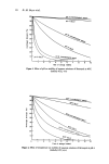
38 B. E. Johnson to uvr in Xeroderma Pigrnentosum differ from those of normal skin, the abnor- mality is not simply a magnification of the normal response and the inference that uvr erythema is mediated through damage to DNA cannot be drawn. Similarly, the vascular reactions in the photosensitised response of 8-methoxypsoralen, almost certainly result- ing from damage to DNA, differ from the normal uvr erythema (75, 76). At the present time, there would appear to be no involvement of DNA in the uvr induced lesions of photodermatoses such as polymorphic light eruption and actinic reticuloid (77). PHARMACOLOGY Ultraviolet erythema has come to be regarded as a typical inflammatory state (78, 79) although the degree of leucocyte migration is significantly less than in other forms of inflammation. Lewis (80) likened sunburn to the reaction obtained with histamine and suggested that the famous 'H' substance was the mediator involved. Histamine depletion and anti-histamine administration prior to irradiation fail to modify the uv-erythema however (81, 82). Serotonin may have a role in rat skin reaction to uvr (83) but does not appear to be involved in human erythema (84). Increased concentrations of vasodilator substances have been detected in blood and skin of uv-irradiated animals (1) but the kinins observed by Epstein and Winkelmann (85) in the perfusate of irradiated human skin are apparently not involved in the delayed erythema reaction. Greaves and Sonder- gaard (86) reported the presence of a prostaglandin-like substance in dermal perfusates of uv-irradiated human skin, the time course of its appearance approximating that of the erythema. Mathur and Gandhi (87) described an increase in prostaglandin content of rat skin after uv-irradiation and indomethacin, a potent inhibitor of prostaglandin synthesis, appears to prevent the development of uvr erythema (88, 89, 90). Greaves and his co- workers have recently shown that Prostaglandin E2 is the major active constituent of uv-irradiated skin, but the site of its derivation has not been ascertained. If it is produced mainly in the epidermis, either as a result of photochemical changes in cellular fatty acids or by a process initiated by lysosomal enzymes, it may well be the mythical, dif- fusible mediator beloved of this field and so elegantly discussed by Leun (13). CHRONIC EFFECTS The long-term effects of repeated exposures to high intensity sunlight have been well reviewed by Blum (91), Epstein (92) and Urbach, Epstein and Forbes (93) a good sum- mary is presented in the book of Magnus (4). The major change observed in the exposed skin of lightly pigmented peoples is an accelerated ageing. The skin loses its natural elasticity, there is marked epidermal atrophy and increased amounts of mucopolysac- charides are found in the dermis, along with deposits of an elastotic material. Focal, benign abnormalities of keratinisation develop to form small, crusty lesions called solar keratoses and these may be succeeded by basal and squamous cell carcinomas. Evidence for the role of solar uvr in skin carcinogenesis in humans is entriely epidemiologic but convincing. The majority of skin cancer, where no other direct carcinogenic influence is known, is found in the sun-exposed skin of lightly pigmented people and is rare in Negroids. There is an approximate correlation between the incidence of skin cancer and the degree of insolation in similar populations living in different areas and turnours develop mostly in areas of the skin most directly exposed to sunlight. This epidemio- logic evidence is supported by data from numerous studies with experimental animals.

Sunburn and mechanisms of protection 39 The carcinogenic action of uvr appears to derive from both the mutagenic effects and the hyperplasia inducing action. The uvr wavelength region involved is basically similar to that for uv-erythema but may well differ in detail and there is little reason to suppose that the mechanisms involved in uvr carcinogenesis are simply those in uv-erythema pro- duetion repeated over a long term. PROTECTION The endogenous mechanisms for protection against the effects of uvr on sktn may operate in three distinct ways. In the first place, uvr damage in the skin may be rapidly repaired: the lack of repair is held responsible for the increased sensitivity seen in Xeroderma Pig- mentosum. Secondly, physiological processes may be present by which the energy of excited state molecules or the reactive oxidation products of uv-irradiation may be quenched or harmlessly dissipated. Natural anti-oxidants such as alpha-tocopherol may act in this way. However, the natural carotenoids do not appear to be effective in the normal sunburn reaction (94). The third and most obvious mechanism involves the attenuation of the incident radiation between the skin surface and the molecular target in the skin. HORNY LAYER In lightly pigmented skin, the major contribution to this attenuation comes from the horny layer of the epidermis. The multi-faceted surfaces present in this dead cell layer produce considerable reflection and scattering and the dense protein content provides a filtering effect by absorption. The importance of this attenuation is demonstrated by the difficulty experienced in producing uv-erythema in palmar or plantar skin, by the in- crease in intensity of erythema obtained in skin from which the horny layer has been removed, and by the fact that in the amelanotic skin of Vitiligo and Albinism thickening of the horny layer affords some protection against uvr damage. Bachem (23) showed that only some 15•o of incident 280-nm radiation penetrated a 0.08-mm thickness of fair- skinned horny layer. At 300 nm, a value of 34•o was obtained, while 80•o of 400-nm radiation penetrated to the Malpighian layer. These results are typical of many obtained in studies designed to assess the protective function of the horny layer or to determine the anatomical site of uvr action in skin. Miescher (95) showed that the penetration of 297-320 nm radiation could be described as an exponential extinction with a half-value layer of about 9 microns. UROCANIC ACID The role of urocanic acid as a natural sunscreen for human skin remains debatable (96, 97, 98, 99, 100). This metabolite of histidine, found in human sweat and at higher con- centrations in the epidermis, has an absorption spectrum which extends into the sunburn uvr. In unexposed skin, it is present mostly in the cis form, uv-irradiation resulting in a cis-trans isomerisation. It is this isomerisation with the absorbed energy being dissipated in the reverse direction which may account for any protective role ascribed. The urocanic acid content of epidermis in skin which has been previously exposed to uvr and shows a decreased sensitivity through accommodation, is found to be increased and it may be that the contribution of this protective mechanism to the overall decrease in sensitivity
Purchased for the exclusive use of nofirst nolast (unknown)
From: SCC Media Library & Resource Center (library.scconline.org)