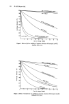
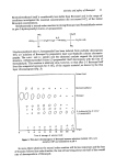
34 B. oe. Johnson for erythema has allowed interesting theoretical treatments of the mechanisms involved in the sunburn reaction (22, 13). Leun (13) concludes that the uvr erythema is brought about by a dermal reaction to all effective wavelengths, the shorter wavelengths being more active, and that a more superficial reaction to the 290-300 nm region results in the formation of a diffusible mediator for erythema, this action being superimposed upon the dermal one to produce a double peak action spectrum. Although most of the incident radiation between 250 and 320 nm is absorbed in the epidermis, at least 1 •o may reach the dermis (23). The penetration of the longer uvr wavelengths is significantly greater than that of the 260-nm region and it appears more likely that the shorter wavelengths would act at a more superficial level. However, the theoretical treatment of uvr erythema published by Leun (13), Ramsay and Challoner (24) notwithstanding, has not been re- pudiated and at this time, the mechanisms suggested fit well with the majority of the experimental data. All the published action spectra show similar features for uvr beyond 290-295 nm, i.e. a rapid fall off in effectiveness so that for all practical purposes, 320 nm and longer wavelength radiation are ineffective, confirming the filtering action of window glass. As the intensity of uvr in natural sunlight falls rapidly from 330 nm to almost zero at 290 nm, the most active wavelength region in natural sunlight will be at a higher value than that shown in Fig. 1. Schulze (25) has computed the most effective wavelength as around 307 nm using the data of a 'standard' erythema action spectrum and the solar radiation measurements of Bener (26). This peak of effectiveness would be moved further into the long-wavelength uvr in geographical locations in which a more rapid fall-off of intensity with decreasing wavelength occurs. As the gradation of severity of reaction increases so markedly with increasing dose at these wavelengths, it is not surprising that a severe sunburn may result from exposure to natural sunlight which appears to be very little beyond a minimal erythema dose. In fair-skinned individuals in Dundee, latitude 56.5N, back skin exposed to 20 min of mid-day June sunlight may show no reaction. A 30-min exposure, however, may resbit in an intense erythema accompanied by tender- ness and oedema. MECHANISMS FOR THE REACTIONS Erythema is the most easily observed component of the sunburn reaction and has there- fore been the most ardently studied. Mild erythema is a manifestation of vasodilatation in the superficial venous plexus of the papillary dermis. More intense erythema, accom- panied by increased skin temperature, reflects arterial and arteriolar dilatation and increased blood flow (24, 27). With very high doses of uvr, vascular stasis may occur (28). Despite the intensive studies of the reaction, the nature and location of the primary molecular target for uvr damage to skin remain unknown. A direct effect of uvr on isolated blood vessels has been described (29) but the immediate contraction observed seems to have little relevance for the delayed onset, persistent erythema observed in irradiated skin. The effects of direct damage to blood vessels in the skin are seen in the photosensitised reac- tion obtained in the disease Erythropoietic Protoporphyria in which excess protopor- phyrin production in the red blood cells leads to abnormal skin reactions to 400-rim radiation. The vascular endothelium is partially destroyed and perivascular deposits of a mucoid substance are laid down as the endothelium is repaired (30). There is no evidence of a similar process in the sunburn reaction and only when excessively high doses of uvr, in particular uv-A, are used, is endothelial damage observed (31).

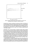
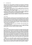
Sunburn and mechanisms of protection 35 HISTOPATHOLOGY The histopathology of the sunburn reaction has been well documented by Rost and Keller (32), Uhlmann (33), Hamperl, Henschke and Schulze (34) and Daniels, Brophy and Lobitz (35). No changes are detected before the appearance oferythema and evidence of damage appears to be restricted to the epidermis. With the appearance of erythema, intracellular oedema in the epidermis and minimal leucocyte migration in the dermis may be observed. By 24 h after the exposure, cellular degeneration is evident in the epidermis which, with minimal erythema, is restricted to individual cells scattered throughout the upper stratum spinosum. With more severe reactions, the number of damaged cells is increased and basal layer cells may be involved. Complete epidermal necrosis is seen in blistering reactions and derreal connective tissue damage may be evident. Epidermal regeneration, as indicated by increased numbers of mitotic figures in the basal layer, occurs by 72 h. The sunburned epidermis is significantly thicker than normal by 6 d, a parakeratotic horny layer being present beneath the still retained, normally appearing original horny layer. The early electron microscope studies of sunburn have been summarised by Nix (36). Cytoplasmic vacuoles appear in the basal and spinous cell layers shortly after exposure but are no longer observed at 72 h. Wilgram et al. (37) have described a decrease in number of keratinosomes in the upper malpighian and granular layer cells as early as 2 h after exposure, but no other changes are observed until 12 h when irregular dense bodies, presumed to be glycogen deposits, appear in the cytoplasm of the basal cells. Increased numbers of thickened tonofibrils are observed in the granular layer at this time. At 72 h, the granular layer is thickened and the cells contain numbers of vacuoles •nd irregular dense bodies. A parakeratotic horny layer, containing well preserved melanin granules and showing evidence of disordered keratinisation, is present between this granular layer and the original horny layer. Nucleolar enlargement, generally accepted as an indication of increased protein synthesis, in spinous cell nuclei, is a major feature of the E.M. picture at 72 h. The isolated, damaged cells, scattered throughout the spinous cell layer and shown by light microscopy to have pyknotic nuclei and shrunken, deeply staining cytoplasm, have been studied at the ultrastructural level (37, 38). These so-called sunburn cells (39) the most specific feature of moderately sunburned epidermis, may be dyskeratotic cells, pushed into premature, and therefore abnormal keratinisation. Alternatively, they may be the epidermal equivalent of the general phenomenon known as 'apoptosis' or indivi- dual cell shrinkage necrosis (40, 41) by which damaged cells are removed from the general population and a stable cell population is maintained. Histochemical studies of sunburn have mainly demonstrated secondary effects and have not been useful in determining the primary site for uvr damage in skin (42). Changes in enzyme activity generally and glycogen deposition (35) appear to be secondary events and decreased nuclear staining in epidermal cells of uv-irradiated skin is not evident until some 24 h after exposure (34, 43) and may represent a stain dilution effect as a result of nuclear swelling, rather than any photochemically induced alteration in DNA struc- ture. There are exceptions to this general rule however. Autoradiographic studies have shown a uvr induced inhibition of DNA, RNA and protein synthesis in skin within the first hour after exposure (44). It may be inferred from these results that DNA and RNA are directly affected in skin by uv-irradiation in vivo. An earlier study (45) had demon- strated 'light labelling' with 3H-thymidine in skin irradiated in vivo, an indication of the
Purchased for the exclusive use of nofirst nolast (unknown)
From: SCC Media Library & Resource Center (library.scconline.org)