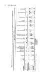
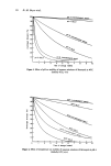
32 B. E. Johnson Blistering may occur in which case, any developing suntan will be lost in the blistered area. Even with a mild sunburn, the outer layers of the stratum corneum tend to be sloughed in sheets rather than as the normal minute scales. When larger areas of the body are exposed, the severe local reactions may be accompanied by nausea, headaches and muscle weakness and medical treatment may be necessary. In already tanned skin, in Asian caucasoids and in the less heavily pigmented negroids, the intensity of the inflammation produced by a given exposure dose is less than that in fair-skinned peoples and even in the latter, slight variations in degree of pigmentation produce differences in the intensity of reaction to sunlight (9). In pigmented skin, an immediate hyperpig- menration can be seen in exposed areas, a phenomenon known as immediate pigment darkening (IPD) which appears to result from an oxidation reaction in melanin already present in the outer layers of the skin but in a bleached form. WAVELENGTH DEPENDENCE Neither infrared radiation (beyond 700 nm) nor visible (390-700 nm) produces the characteristic sunburn reaction. Window glass, acting as a cut-off filter at 315-320 nm, prevents the reaction and this sets the upper limit for effective radiation at around 315 nm. The lower limit is set by the absorption of short wavelength ultraviolet radiation (uvr) by stratospheric ozone, the lowest value of wavelength recorded at the earth's surface being around 285 nm. It would seem that natural sunburn is produced by uvr between 280 and 315 nm, a minute fraction of the total solar spectrum. Because this wavelength region is so important for photobiology, the more conventional partition of the uv spectrum into far-uv (180-290 nm), and near-uv (290-390 nm) has been modified (10) into uv-A (315-390 nm), uv-B (280-315 nm) and uv-C (180-280 nm) a classification which has found considerable usage in dermatological literature. Artificial sources are essential for controlled studies of sunburn and other biologic effects of uvr. Low pressure mercury arcs emit principally the 254-nm line and this uv-C radiation does produce a sunburn-like reaction in human skin. There may be an additive or augmenting action of uv-A on the natural sunburn reaction (11, 12) and uv-B, uv-A and visible radiation are separately or collectively involved in the abnormal reactions seen in the various photodermatoses. There are significant differences between the erythema produced by exposure to uv-C, uv-B and natural sunlight (7, 8, 13). The uv-C erythema is less intense and has a shorter duration than that produced by uv-B. While intense irritation and epidermal damage may be caused, the severe lesions, involving damage to the dermis, which may develop with uv-B irradiation cannot be elicited with uv-C. On the other hand, the uv-B erythema has a shorter duration than that produced by sunlight and the latent period for its develop- ment is relatively prolonged. Thus the natural sunburn response is not necessarily pro- duced by exposure to a narrow band of uvr wavelengths and the reports of Hausser (14) that uv-A erythema, although requiring a very high exposure dose, may be observed very soon after the exposure and persists, and of Willis, Kligman and Epstein (11) and Parrish et al. (12) that the dose requirement for uv-B erythema may be reduced by exposure to uv-A, appear to have considerable relevance to the problem of the natural sunburn. Detailed studies of the wavelength dependency for uvr erythema began with the work of Hausser and Vahle (15). An action spectrum for the just perceptible erythema in upper back skin of lightly pigmented individuals is shown in Fig. I. This was plotted from data obtained by Dr John Anderson and myself at Dundee, using a small grating irradiation

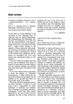
Sunburn and mechanisms of protection 33 Figure 1. Action spectrum for just perceptible erythema in fair-skinned human subjects back skin, readings at 7-8 and 24 h. The 100K level at 260 nm represents a mean MED of 4.3 mJcm -•, standard deviation 1.4. 100, 250 260 2,0 2[0 go ' 310 WAVELENGTH (rim) monochromator (Rank-Hilger, D330 with D335 grating) with a 450 W Xenon arc source (Osram, XBO 450 W). The half-maximum bandwidth used was 2.6 nm, giving minimal dispersion around the selected wavelength. Readings of the reactions were made at 7-8 h and 24 h after the exposures. The minimum dose required to produce a reaction, whether observed at 7-8 h or 24 h, was taken as the minimal erythema dose (MED). The action spectrum is plotted from the reciprocal of the MED at each wavelength and is a relative effectiveness curve. It is typical of the various action spectra published in the past (3) and resembles those of Cripps and Ramsay (16) and Nakayama et al. (17). Problems have arisen with regard to the shape of the action spectrum in the wavelength region below 285 nm which were foreshadowed in the work of Hausser and Vahle (18). In the action spectra published before that of Everett, Olson and Sayre (19) the wavelength region around 260 nm, although shown to have a peak of activity, was less effective than the 290-nm region. Where the reaction observed is a minimally perceptible erythema, it is obvious from Fig. I that the shorter wavelengths are, in fact, more effective. Moreover, the trough in the action spectrum at 280 nm is much deeper in the earlier publications. Explanations for these discrepancies were proposed by Everett et al. (19) and by Berger, Urbach and Davies (20) and these are elegantly discussed by Leun (21). A minimal erythema produced by 260-nm radiation may develop by 8 h but fade before 24 h. Thus, for erythema readings at 24 h only, a higher dose is required for the shorter wavelengths' reactions and the activity of this wavelength region will appear to be relatively low. More- over, the gradation of erythema redness produced by the two wavelength regions with increasing dose is different. While the intensity of the 260-rim reaction increases slowly and reaches a low plateau level, that for longer wavelengths increases rapidly to a much higher plateau level. If some degree of erythema other than the minimally perceptible is chosen as the end point for plotting an action spectrum, as was the case in the earlier studies, the longer wavelengths will again appear to be more effective. The trough in the action spectrum at 280 nm has always been ascribed to the filtering effects of the stratum corneum. This may be true as the trough is deeper in action spectra obtained with forearm skin with a relatively thick corneum, than in those obtained with back skin. The prolonged arguments about the effects of uvr wavelengths not present in natural sunlight may appear superfluous. However, these shorter wavelengths are a prominent part of uvr photobiology, particularly in the investigations of cellular mechanisms of uvr damage. Nucleic acids have major absorption peaks in this wavelength region as do proteins through the aromatic amino acid constituents. Also, the total action spectrum
Purchased for the exclusive use of nofirst nolast (unknown)
From: SCC Media Library & Resource Center (library.scconline.org)