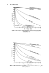
40 B. E. Johnson of accommodated skin is greater than that of the protein constituents of the horny layer. MELANIN The major contributory factor in the natural protection of human skin against uvr is the complex, indole type polymer called melanin. The relationship between sunlight and melanin pigmentation has been most recently reviewed by Pathak et al. (9), and Quevedo et al. (101), Szabo (102), and Pathak (103) present good coverage of the subject matter as known at that time. The development of a suntan in normally lightly pigmented skin, resulting from a uvr-stimulated redistribution of preformed melanosomes in the epi- dermis and the tyrosinase controlled production of melanised melanosomes in the melanocytes with subsequent transfer to and distribution in the keratinocytes of the hyperplastic epidermis, obviously affords protection against further uvr exposure. The genetically controlled variation in number, form and distribution of melanosomes, mainly responsible for racial variation in skin colour, results also in variations in the skin responses to uvr. In fair-skinned Celtic peoples, Albinos and in vitiliginous skin, where little or no melanin is present, the skin reactions are severe. As the degree of melanisation increases, so the intensity of skin reactions decreases and in deeply pig- mented Negroids and Australoids, it is difficult to elicit reactions other than the IPD and skin cancer due to uvr does not occur. It is thought that the racial variation in human skin colour may represent an evolutionary compromise between the need to protect against the carcinogenic action of uvr and the requirement of photochemically-produced vitamin D in the viable layers of the epidermis (104, 105). The transmission of 280-320 nm radiation through negro horny layer is significantly less than through that from white skin, but there is no significant difference in the thickness of this layer (106, 107). The only difference appears to be the presence of melanin as demonstrated by Kligman (108). The mechanisms by which melanin exerts its protective function apparently vary. It may act as a neutral density filter in the outermost layers of the epidermis where it is present in a disperse, amorphous form. The absorbed energy is dissipated harmlessly as heat. Moreover, in this form it acts as an excellent scattering screen for the wavelengths involved. The scattering produced by the larger melanin granules, usually observed in a supranuclear distribution in the basal and suprabasal layers, is thought to involve a high degree of forward direction, and if this is so the function of the superanuclear caps in protecting the germinal DNA is a little difficult to accept. Stable free radicals are present in the melanin polymer and may act as electron traps in a uvr-excited system. However, the spatial relationship between the potentially susceptible biological substrate, for instance DNA, and the melanin would have to be rather more intimate than it appears in electron microscope pictures for this protective mechanism to function efficiently. It is also thought that melanin may act as a semi-conductor material, in this way diverting electron energy away from susceptible biological material. The exact mechanisms involved in melanin protection against uvr damage, apart from the action as a filter, are obviously not as yet worked out, but it would seem that they do exist and are effective. CONCLUSION In summary, the reactions of human skin to the uvr component of solar radiation may vary according to the intensity and wavelength of the incident radiation from the mildest

Sunburn and mechanisms of protection 41 form of inflammation to severe blistering and necrosis. With repeated exposures, pre- mature ageing and cancer of the skin may develop. The skin is probably a multi-target system for uvr effects and natural protection against these effects resides predominantly in the melanin content of the skin. REFERENCES 1 Blum, H. F. Physiological effects of sunlight on man. Physiol. Rev. 25 483 (1945). 2 Kimmig, J. and Wiskemann, A. Handbuch der Haut- und Geschlechtskrankheiten, Erganzungswerk vol. 5, part 2, 1021 (1959) Springer, Berlin. 3 Johnson, B. E., Daniels, F., Jr. and Magnus, I. A. Response of human skin to ultraviolet light. In: Giese, A. C. Photophysiology vol. 4, 139 (1968) Academic Press, London. 4 Magnus, I. A. Dermatological Photobiology: Clinical and Experimental Aspects (1976) Blackwell Scientific Publications, Oxford. 5 Urbach, F. The Biologic Effects of Ultraviolet Radiation (with emphasis on the skin) (1969) Pergamon Press, Oxford. 6 Fitzpatrick, T. B. Sunlight and Man normal and abnormal photobiologic responses (1974) University of Tokyo Press. 7 Buettner, K. J. K. The effects of natural sunlight on human skin. In: Urbach, F. The Biologic Effects of Ultraviolet Radiation, 237 (1969) Pergamon Press, Oxford. 8 Breit, R. and Kligman, A. Measurement of erythemal and pigmentary responses to ultraviolet radiation. In: Urbach, F. The Biologic Effects of Ultraviolet Radiation 267 (1969) Pergamon Press, Oxford. 9 Pathak, M. A., Jimbow, K., Szabo, G. and Fitzpatrick, T. B. Sunlight and melanin pigmentation. In: Smith, K. C. Photochemical and Photobiological Reviews vol. 1,211 (1976) Plenum Press, New Yolk. 10 Coblentz, W. Ultraviolet radiation useful for therapeutic purposes specification of minimum in- tensity or radiant flux second communication. J. Am. Med. Assoc. 99 125 (1932). 11 Willis, I., Kligrnan, A. and Epstein, J. H. Effects of long ultraviolet rays on human skin: photo- protective or photoaugmentative. J. Invest. Dermatol. 59 416 (1972). 12 Parrish, J. A., Ying, C. Y., Pathak, M. A. and Fitzpatrick, T. B. Erythemogenic properties of long- wave ultraviolet light. In: Fitzpatrick, T. B. Sunlight and Man 131 (1974) University of Tokyo Press. 13 Leun, J. C. v. d. Ultraviolet erythema. A study of diffusion processes in human skin. Thesis (1966) University of Utrecht. 14 Hausser, I. fiber spezifische Wirkungen des langwelligen Ultravioletten Lichts auf die menschliche Haut. Strahlentherapie 62 315 (1938). 15 Hausser, K. W. and Vahle, W. Die Abhangigkeit des lichterythems und der Pigrnentbildung von der Schwingungszahl (Wellenlange) der erregenden Strahlung. Strahlentherapie 13 41 (1922). 16 Cripps, D. J. and Ramsay, C. A. Ultraviolet action spectrum with a prism-grating monochromator. Brit. J. Dermatol. 82 584 (1970). 17 Nakayama, Y., Fukuda, M., Yano, K., Morikawa, F. and Mizuno, N. Studies of erythema action spectrum with a monochromator. Jap. J. Dermatol. Ser. A 81 921 (1971). 18 Hausser, K. W. and Vahle, W. Sonnenbrand und Sonnenbraunung. Wiss. Veroff. Siemens-Werke 6 101 (1927). 19 Everett, M. A., Olson, R. L. and Sayre, R. M. Ultraviolet erythema Arch. Dermatol. 92 713 (1965). 20 Berger, D., Urbach, F. and Davies, R. E. The action spectrum of erythema induced by ultraviolet radiation. In: Jadassohn, W. and Schirren, C. G. XIII Cbngressus Internationalis Dermatologiae vol. 2, 1112 (1968) Springer, Berlin. 21 Leun, J. C. v. d. On the action spectrum of ultraviolet erythema. In: Gallo, U. and Santamaria, L. Research Progress in Organic, Biological and Medicinal Chemistry vol. 3, part 2, 711 (1972) North Holland. 22 Mitchell, J. S. Origin of erythema curve and pharmacological action of ultraviolet radiation. Proc. R. Soc. Lonabn Series B 126, 241 (1938). 23 Bachem, A. Die Lichtdurchdringung der menschlichen Haut. Strahlentherapie 39 30 (1930). 24 Ramsay, C. A. and Challoner, A. V. J. Vascular changes in human skin after ultraviolet irradiation. J. Dermatol. 94 487 (1976).
Purchased for the exclusive use of nofirst nolast (unknown)
From: SCC Media Library & Resource Center (library.scconline.org)