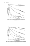
Sunburn and mechanisms of protection 35 HISTOPATHOLOGY The histopathology of the sunburn reaction has been well documented by Rost and Keller (32), Uhlmann (33), Hamperl, Henschke and Schulze (34) and Daniels, Brophy and Lobitz (35). No changes are detected before the appearance oferythema and evidence of damage appears to be restricted to the epidermis. With the appearance of erythema, intracellular oedema in the epidermis and minimal leucocyte migration in the dermis may be observed. By 24 h after the exposure, cellular degeneration is evident in the epidermis which, with minimal erythema, is restricted to individual cells scattered throughout the upper stratum spinosum. With more severe reactions, the number of damaged cells is increased and basal layer cells may be involved. Complete epidermal necrosis is seen in blistering reactions and derreal connective tissue damage may be evident. Epidermal regeneration, as indicated by increased numbers of mitotic figures in the basal layer, occurs by 72 h. The sunburned epidermis is significantly thicker than normal by 6 d, a parakeratotic horny layer being present beneath the still retained, normally appearing original horny layer. The early electron microscope studies of sunburn have been summarised by Nix (36). Cytoplasmic vacuoles appear in the basal and spinous cell layers shortly after exposure but are no longer observed at 72 h. Wilgram et al. (37) have described a decrease in number of keratinosomes in the upper malpighian and granular layer cells as early as 2 h after exposure, but no other changes are observed until 12 h when irregular dense bodies, presumed to be glycogen deposits, appear in the cytoplasm of the basal cells. Increased numbers of thickened tonofibrils are observed in the granular layer at this time. At 72 h, the granular layer is thickened and the cells contain numbers of vacuoles •nd irregular dense bodies. A parakeratotic horny layer, containing well preserved melanin granules and showing evidence of disordered keratinisation, is present between this granular layer and the original horny layer. Nucleolar enlargement, generally accepted as an indication of increased protein synthesis, in spinous cell nuclei, is a major feature of the E.M. picture at 72 h. The isolated, damaged cells, scattered throughout the spinous cell layer and shown by light microscopy to have pyknotic nuclei and shrunken, deeply staining cytoplasm, have been studied at the ultrastructural level (37, 38). These so-called sunburn cells (39) the most specific feature of moderately sunburned epidermis, may be dyskeratotic cells, pushed into premature, and therefore abnormal keratinisation. Alternatively, they may be the epidermal equivalent of the general phenomenon known as 'apoptosis' or indivi- dual cell shrinkage necrosis (40, 41) by which damaged cells are removed from the general population and a stable cell population is maintained. Histochemical studies of sunburn have mainly demonstrated secondary effects and have not been useful in determining the primary site for uvr damage in skin (42). Changes in enzyme activity generally and glycogen deposition (35) appear to be secondary events and decreased nuclear staining in epidermal cells of uv-irradiated skin is not evident until some 24 h after exposure (34, 43) and may represent a stain dilution effect as a result of nuclear swelling, rather than any photochemically induced alteration in DNA struc- ture. There are exceptions to this general rule however. Autoradiographic studies have shown a uvr induced inhibition of DNA, RNA and protein synthesis in skin within the first hour after exposure (44). It may be inferred from these results that DNA and RNA are directly affected in skin by uv-irradiation in vivo. An earlier study (45) had demon- strated 'light labelling' with 3H-thymidine in skin irradiated in vivo, an indication of the

36 B. E. Johnson repair process by which uvr-induced thymine dimers are removed from DNA (46). This is perhaps the first report to show that DNA is a direct target for skin reactions to uvr and it may be inferred from the results that thymine dimers are the molecular lesions involved. In addition, fluorescent antibody techniques, using antibodies raised against uv-irradiated native DNA, have been used to demonstrate damage in DNA of skin irradiated in vivo (47, 48). As thymine dimers are the most prominent of the DNA photo- products, these studies can be interpreted as demonstrating, although again indirectly, that thymine dimers are formed in skin irradiated in vivo. Wavelength dependency studies of this reaction show that it is restricted to the sunburn region of the uv-spectrum (49). Changes in intensity and distribution of the staining reaction for acid phosphatase activity in uv-irradiated skin have been interpreted as evidence that the lysosome mem- brane is a primary target for uvr damage and that lysosome enzymes released as a con- sequence of this damage may be involved in the secondary processes of sunburn (39, 50). The decrease in keratinosome number reported by Wilgram et al. (37) appears to support this hypothesis, but Honigsmann et al. (51) could find no evidence of early lysosome membrane disruption in epidermal cells of uv-irradiated skin using ultra- structural techniques. BIOPHYSICS AND BIOCHEMISTRY A specific free radical signal is produced in lightly pigmented skin on exposure to sun- burning uvr (52) but the chemical nature of this excited molecular state has not been identified. Lipid peroxide, possibly derived from free radical induced reactions in un- saturated fatty acids and relatively long lived, has been identified in uv-irradiated skin (53, 54). Lipid peroxides have considerable biological activity which includes the inhi- bition of respiration and glycolysis, oxidation of sulphydryl groups and destruction of anti-oxidants such as vitamin E. Moreover, lysosome membranes are particularly sus- ceptible to labilisation by lipid peroxides which have been incriminated in cell necrosis in carbon tetrachloride damaged liver and in inflammation of the intestinal mucosa. Peroxides isolated from uv-irradiated skin have been shown to be potent carcinogens (55) but the role, if any, of these compounds in uvr inflammation has still to be defined. Under ideal conditions, the action spectrum for a photochemical reaction should fit the absorption spectrum for the molecule involved. In the photosensitive porphyrias the action spectrum for the abnormal response fits the soret band absorption characteristic of the porphyrins. The finding of a peak of erythemal activity at around 260 nm and general activity at longer wavelengths has focused attention on nucleic acids and proteins as the primary target molecules for skin reactions to uvr. However, skin is so heterogeneous a medium, uvr energy may be absorbed and dissipated as harmless quantities of heat or as fluorescence, that such a treatment is hopeful if not naive. The action spectrum presented by Everett et al. (19) might well be interpreted as representing an absorbance in un- saturated fatty acids to initiate the reaction, deviations from the shape of a smooth absorption curve being explained in terms of the filtering effects of the horny layer. Nonetheless, DNA appears to be the major molecular target for uvr lethality and muta- genesis in bacteria and mammalian cells in culture (56, 57, 58). Although DNA-protein binding may be involved in these effects, the most prominent and contributory molecular lesion is the cyclobutane type dimer formed between adjacent thymines on a single strand of the DNA double helix. Inhibition of DNA synthesis, cell death and mutation may result directly from the presence of thymine dimers or indirectly through faulty
Purchased for the exclusive use of nofirst nolast (unknown)
From: SCC Media Library & Resource Center (library.scconline.org)