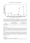
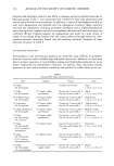
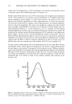
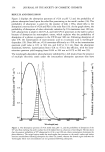
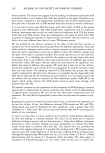
152 JOURNAL OF THE SOCIETY OF COSMETIC CHEMISTS could occur (2). Subsequently, t-UA was thought to be harmless and therefore used as a sunscreen and/or skin-conditioning agent in cosmetics (3). Further research has made such uses of UA controversial. For example, upon absorption of UV, t-UA isomerizes to cis-urocanic acid (c-UA) (Scheme 1) (4). Both isomers have similar absorption spectra (Figure 1), which were found to be similar to the action spectrum for immune suppression of contact hypersensitivity (5). Since this discovery, c-UA has been determined to suppress contact hypersensitivity and delayed hypersen- sitivity, reduce the Langerhans cell count in the epidermis, prolong skin graft survival time, and affect natural killer cell activity (6-12). Because c-UA has shown such im- munomodulatory behavior, it is now postulated to act as a mediator for UV-induced immunosuppression however, this is not proven because the mechanism by which UA modulates the immune system following absorption of UV radiation is not definitively known. Hence, the use of UA in topical formulations remains controversial. The U.S. Food and Drug Administration does not list UA as one of its restricted or prohibited ingredients to cosmetics, but use of UA in the United States is extremely rare. A request conducted by the FDA in 1991 for volunteer information on the use of UA in the U.S. reflected that only one manufacturer sells products with UA as an ingredient (3). To gain a better understanding of how the photochemistry of UA could affect the skin and therefore either caution against or promote the use of UA in topical formulations, we have begun a spectroscopic investigation of the molecule in vitro. Different spectros- copies were employed to investigate the unusual photochemical behavior t-UA exhibits. We have recently reported a photochemical study of UA as a function of pH and isomer (13). Here, we summarize our results from this study and comment on how the pho- tochemical data is of interest to those in the cosmetic industry who continue to use urocanic acid. 15xl 03 T..•, 10 5 0 240 260 280 300 320 340 Wavelength (nm) Figure 1. Absorption spectra of trans- and cis-urocanic acid (pH 7.2). Both isomers absorb in the UV-B, with c-UA (dashed line) having a smaller molar extinction coefficient (½ = 13,600 at 278 nm) relative to the naturally occurring t-UA isomer (solid line, ½ = 18,800 at 278 nm).

PHOTOCHEMISTRY OF UROCANIC ACID 153 MATERIALS AND METHODS REAGENTS AND SAMPLE PREPARATION All experiments were accomplished in a quartz cuvette using 10 -3 M urocanic acid solutions that were buffered. trans-urocanic acid was purchased from Aldrich (Milwau- kee, WI) and used without further purification. Bromocresol purple was purchased from Fischer. All solutions were buffered in either 0.1 M acetate buffer, pH 5.6, the average pH of the stratum corneum and sweat (14), or 0.1 M potassium phosphate, pH 7.2, the average pH of living cells. c-UA was isolated following the procedure of Anglin and Everett (15). ABSORPTION SPECTRA AND PROBABILITY OF UV ABSORPTION Absorption spectra were recorded on an HP 8245 A diode array spectrophotometer. The probability of absorption of solar radiation was determined from the product of the extinction coefficient of t-UA at pH 7.2 and the solar flux penetrating to the earth's surface at 30 ø (16). PHOTOACOUSTIC CALORIMETRY Experiment and theory (17). In this paper, we do not present our photoacoustic (a.k.a. ultrasonic) data, as it has been published previously (13) however, because we refer to the data in the following discussion, we give a brief description of the technique and the information it provides for those who are unfamiliar with this type of spectroscopy. Photoacoustic calorimetry provides fundamental energetic information on a chromo- phore. As a result, this technique can be used as a spectroscopic tool to determine if the absorbing molecule dissipates the absorbed energy through a singlet state or through potentially photosensitizing or photoreactive triplet state(s), with radical, biradical, or reaction intermediates following absorption into the excited state (18). The latter are of concern in photobiology because reactive intermediates can lead to damage on the cellular level. The technique works as follows: Following absorption of light by a molecule, the energy absorbed can be released through different pathways (both radiative and nonradiative) or retained by the molecule in intermediate state(s). The total energy absorbed must be equal to the sum of the energy released back to the solvent and the energy retained by the molecule in intermediate state(s). The energy released by the molecule to the surrounding solvent is determined as an ultrasonic wave by a transducer connected to the cuvette containing a solution of the molecule and the solvent. The timescale of the energy dissipation processes measured is determined, in part, by the frequency response of the transducer. In our experiment, the heat released on a timescale of 10 -9 s or faster can be recorded. By comparing the amplitude of the ultrasonic wave of the molecule (tram-urocanic acid) to a reference (bromocresol purple) known to release all of the absorbed energy to the solvent, the amount of energy retained by the molecule can be determined. If any energy is retained by the molecule, it is then determined that the molecule forms a long-lived intermediate state, which could be potentially reactive as discussed above.
Purchased for the exclusive use of nofirst nolast (unknown)
From: SCC Media Library & Resource Center (library.scconline.org)