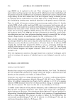
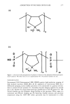
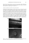
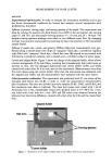
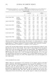
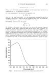
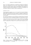
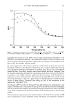
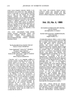
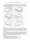
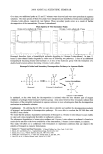
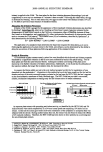
278 JOURNAL OF COSMETIC SCIENCE hydrated state for long periods of time. Resolution is comparable to a conventional SEM (-5 nm with a tungsten gun). Techniques like energy dispersive spectroscopy (EDS) are routinely employed, but there are special considerations due to the gas in the chamber (13,14). There is little literature on ESEM applications to human hair, but related studies include the study of wool (15) and the development of detergents and personal products (16). In this work a Philips-Electroscan ESEM 2020 was used with a PGT IMIX EDS system. Hair was clamped in a special metal stub designed to hold hair strands fiat and to allow reproducible positioning in the ESEM. In this way, individual strands could be exam- ined before and after treatment with conditioning polymers. The stub was temperature- controlled via a Peltier stage. Specimens were typically imaged at -5øC and chamber pressure -5 Torr to maintain approximately 100% RH. X-ray photoelectron spectroscopy (XPS). XPS analyzes the near-surface of materials with analysis depths up to 50 •. It can give quantitative chemical information as well as oxidation and structural environments on all elements apart from hydrogen and helium. Soft X-rays, of energy hv, excite valence electrons from valence and core orbitals of surface atoms. This is shown in Figure 2. The kinetic energy of the ejected electron, EK, is measured by an electron energy analyzer. These photoelectrons have energies according to the relationship (17) E B -- hv - E K - where (I) is the work function of the spectrometer. E B is the binding energy of the photoelectron to the parent atom, and this can be calculated from the other measured and known values. XPS spectra are traditionally plotted as EB vs photoelectron intensity. XPS peaks can then be identified using tabulated binding energy values from XPS handbooks yielding information on chemical composition and bonding environments. Tresses of untreated and polymer-treated hair were clamped into separate metal stages such that a mesh of hair at least 5 mm by 5 mm could be studied. XPS spectra were acquired using a Fisons ESCAscope photoelectron spectrometer with dual anode X-ray 2P3/2 2Pl/2 2S llllllllllll •llllmml Vacuum level core Initial state Final state E K E B hD Figure 2. Emission of a ls photoelectron after X-ray beam of energy hv has interacted with atom. E B is electron binding energy and E K the kinetic energy after leaving vacuum level.

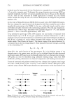
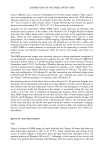
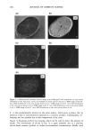
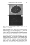
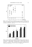
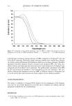
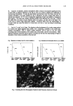
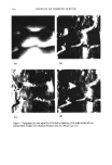
ADSORPTION OF POLYMER ONTO HAIR 279 sources (Mg/A1) and a concentric hemispherical electron energy analyzer. Data acquisi- tion and manipulation was carried out using microprocessor units with VGS software. During acquisition of spectra the pressure in the main chamber was maintained at 8 x 10 -9 torr to ensure a clean sample surface. Charge referencing was carried out against adventitous hydrocarbon (Cls 284.6 eV) (17) from pump oil contamination. Secondary ion mass s[•ectrometry (SI&IS). SIMS allows a mass spectrum of elemental and molecular species present in the surface to be obtained (18). A highly focused ion beam penetrates the surface under study, sputtering atoms and ions in the uppermost surface layer, which are then analyzed according to their mass/charge (m/z) ratio by a mass spectrometer. In a manner similar to SEM imaging, high-resolution SIMS images were obtained by raster scanning the beam over the desired area on the hair's surface and detecting locations of sputtered ions having a prefixed m/z value. However, in contrast to XPS, SIMS is a surface-destructive technique due to the sputtering or erosion of the surface atoms, and therefore acquisition times for spectra and images were kept to a minimum. The SIMS spectra and images were obtained using an in-house instrument comprised of an electronically variable aperture-type gallium ion gun (FEI SD Gallium LMIS EVA focusing column) fitted to a double-focusing magnetic-sector mass analyzer (Vacuum Generators model 7035). An Everhart-Thornley electron detector was used for acquisi- tion of secondary electron images. For secondary ion analysis, a lnA, 25keV impact Ga + microbeam was used, which allowed an image resolution of 300 nm. The upper mass limit was configured at 100 daltons (D), and the instrument was calibrated using values of 68.926 and 70.925 D for the backscattered Ga + ions. Analysis was carried out using the "Dayta" software package (19) running under Windows 95. For obtaining spectra of the hair's outer surface (epicuticle), 10-mm hair lengths were mounted over an aluminium stub using silver dag adhesive at either ends of the hair. It was essential that the hair made good contact with the stub throughout its length, and so an aluminium mesh was placed over the sample to essentially clamp the hair and enable it to lie flat. Due to problems of imaging hair lengths, which will be outlined later, SIMS images were obtained on hair cross sections. Here 10-mm-length samples of both N- and P-Merquat-100-treated hair were embedded in separate blocks of Agar 100 epoxy resin and then microtomed with a diamond knife. Due to the insulating nature of the resin and hair, the blocks were gold-coated for analysis to eliminate charging. Careful coating of the hair under high vacuum would not affect the elemental distri- bution, and the metal layer was etched away before analysis using the lnA 25keV Ga + microbeam. RESULTS AND DISCUSSION ESEM Figure 3a shows an ESEM image of virgin bleached hair. Figure 3b shows hair from the same batch after exposure to pH 9.5 at 20.9øC. Some roughening and raising of the cuticle is visible. Figure 3c shows hair from the same batch after treatment with a 3% aqueous solution of Merquat-100 at pH 7. Although a little raising of the cuticle still seems to be present after treatment with Merquat-100, this may be expected, as the hair
Purchased for the exclusive use of nofirst nolast (unknown)
From: SCC Media Library & Resource Center (library.scconline.org)