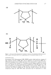
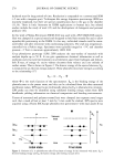
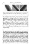
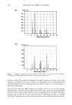
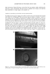
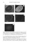
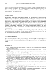
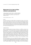
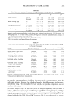
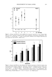
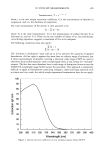
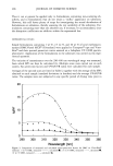
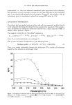
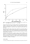
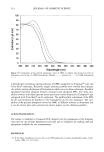
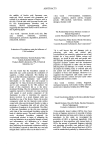
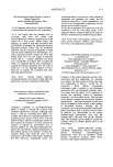
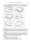
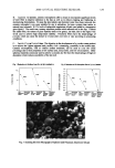
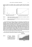
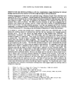
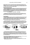
282 JOURNAL OF COSMETIC SCIENCE (a) Counts / a.u. 700 6OO 500 400 3O0 200 100 5 10 5 20 25 30 35 40 45 50 Mass/Charge (Daltons) (b) Counts / a.u. 500 ..... '• ..................................... ', : : 400 300 ............................................':.........j...................... 100 .... 5 10 15 20 25 30 35 40 45 50 Mass/Charge (Daltons) Figure 5. a: Negative ion spectrum of untreated bleached hair. Counts are in arbitrary units. b: Negative ion spectrum of N-Merquat©-100-treated hair. Counts are in arbitrary units. similar to that of the untreated, although the relative amount of chlorine on the hair appears to be higher. The presence of chlorine in the polymer may be responsible for the observed higher count rate, although from this evidence alone it is not yet possible to draw firm conclusions. Elemental and molecular SIMS imaging was initially carried out on the hair lengths, although due to the hair not lying flat on the stub, image quality was poor and much of the contrast in these images is related to the topography of the specimen. Therefore, imaging was carried out on hair cross sections by embedding in resin as detailed previously. Figure 6a shows the microtomed surface of the resin and hairs obtained by

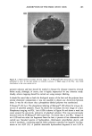
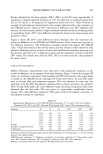
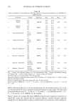
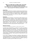
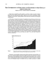
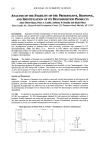
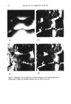
ADSORPTION OF POLYMER ONTO HAIR 283 light microscopy. Figure 6b shows a typical hair that was studied, clearly exhibiting the epicuticle, cortex, and medulla. The cross section is slightly oval in shape, as the hair has been embedded at a slight angle to the normal cut. ELEMENTAL AND MOLECULAR MAPPING OF HAIR CROSS SECTIONS Secondary electron and ion images were taken over the cross section of many hairs treated with either N- or P-Merquat©-100, and typical maps are shown in Figures 7 and 8. It was assumed that the elemental distribution remained constant along the hair during imaging. This was supported by etching a further 20 nm into the hair and re-imaging. It was observed that elemental maps were similar to those acquired on the initial surface. N-Merquat©-100 hair. Figure 7a gives the backscattered electron image of a hair em- bedded in resin. It should be noted that the hair has remained similar in appearance to that taken by the optical microscope, and so it would appear that the effect of ultra-high vacuum on the hair has not affected it too greatly. Figure 7b shows how CN- (26D) mapping can be used to emphasize the biological matrix, and Figure 7c shows how O- sites are primarily located in the outer surface of the hair. Figure 7d indicates that the (a) (b) Figure 6. a: Optical microscope image of microtomed surface of resin and hair to allow flat hair cross- section studies. b: Optical microscope image of a typical hair cross section under study.
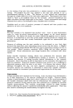
Purchased for the exclusive use of nofirst nolast (unknown)
From: SCC Media Library & Resource Center (library.scconline.org)