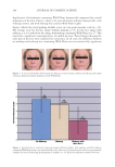
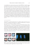
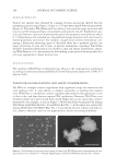
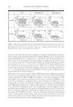
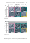
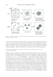
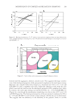
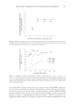
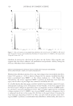
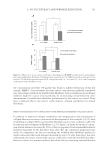
JOURNAL OF COSMETIC SCIENCE 312 INTRODUCTION Aging is now generally regarded as a failure of an organism to repair tissue at the same rate as it is damaged (1,2). The changes occurring during aging are associated with al- terations in skin appearance due to loss of tensile strength, with wrinkle formation, in- creased fragility, and decreased epidermal moisture content. Among these effects, wrinkles are an important indicator of aging and an interesting fi eld in cosmetic dermatology. The appearance of wrinkles is related to an infl exibility of the skin coupled with a slackening of the dermis (3,4). This arises from a reduced production of collagen fi bers accompanied by the degeneration of the surrounding collagenous network (5–7). Resident fi broblasts are intimately involved in the decreased synthesis of collagens such as type I procollagen and an increase in the degradation of collagens. This is through increased collagenases such as matrixmetalloproteinase-1 (MMP-1), which occurs as fi broblasts age (8–10). In addition, aged fi broblasts lose their capacity to adhere and move over collagen fi bers, thereby limiting their ability to reorganize and reorient dermal tissue including collagen fi bers (11–13). For this reason, the improvement of fi broblast activities in terms of dermal tissue biosynthesis and reorganization may be useful for a variety of cosmetic and thera- peutic applications. It has been demonstrated that agents such as all-trans retinol (14–17) and vitamin C (18,19) can stimulate collagen production and suppress MMP activity. In addition, it has been reported that vitamin C and soy peptides can restore the capacity of fi broblasts to realign the collagen fi bers, which in turn improves locomotion, distribution, and adhe- sion of fi broblasts in the collagen matrix (11). These fi ndings indicate that aged-related fi broblast dysfunction is, at least in part, reversible. Thus it would be realistic to develop new agents whose activities are similar to those of retinol- and/or soy proteins that could be applied as cosmetics. In the cosmetics industry, there is demand for multifunctional and effi cacious products based on real innovation and aligned with trends in the cosmetics market. For example, natural products with antioxidant, antityrosinase and antiaging activities have been sought for the treatment of photoaging, unwanted skin pigmentation, and wrinkles. Recently, we found that diethyl either extracts using the heartwood from the Moraceae family, particularly Artocarpus incisus (breadfruit or Sa-kae in Thai) exhibited antioxi- dant activity in a dose-dependent manner using the DPPH assay (20). Furthermore, it has been reported that a crude extract as well as some purifi ed compounds isolated from A. incisus’s heartwood inhibited melanin production (20–22). In the present study, therefore we compared the effects of the extract on some biological functions of fi bro- blasts from nonwrinkled and wrinkled skin that were obtained from a biopsy of skin at the outer corner of the eye. The fi broblast functions assessed included the stimulatory effect on proliferation, production of type I procollagen, and the inhibitory effect on the production of the major collagen-degrading enzyme (MMP-1). In addition, we studied the effect of the extract on the ability of “wrinkled-skin fi broblasts” to reorga- nize collagen fi bers in a collagen lattice. The effect can be measured as a contraction of the collagen lattice, which thus indicates reorganization capacity (23,24). The results obtained from our study fi rst revealed the biological effects A. incisus’s heartwood ex- tract on human wrinkled-skin fi broblasts that were distinct from non-wrinkled skin cells. They also show that A. incisus’s heartwood extract could have potential cosmetic applications.

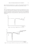
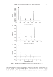
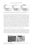
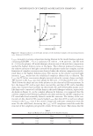
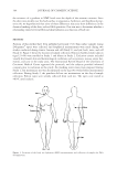
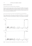
A. INCISUS EXTRACT AND WRINKLE REDUCTION 313 MATERIALS AND METHODS PLANT MATERIAL AND THE EXTRACTION PROCESS The heartwood of A. incisus was collected from Phitsanulok Province, Thailand. The heartwood portion of A. incisus was chipped and dried at 50°C by using a hot-air oven. Then the dried-chipped heartwood was milled into a powder. Five-hundred grams of the A. incisus powder was macerated with 800 ml of diethyl ether (LabScan Asia, Co. Ltd., Bangkok, Thailand) at room temperature for two days, as used previously with slight modifi cation (20). The mixture was fi ltered through a cloth to remove particu- lates, and then the diethyl either was removed by evaporation with a vacuum evapora- tor set at 33°C. The resultant powder was stored in a tight amber glass at −20°C for further studies. QUALITY CONTROL OF THE EXTRACT To control the extract quality of each batch, the content of the artocarpin, a major com- ponent of the A. Incisus heartwood extract, was determined by using isocratic high-per- formance liquid chromatography (HPLC). The artocarpin was provided by Assist Prof. Atawit Somsiri, Faculty of Pharmaceutical Sciences, Naresuan University (25). The HPLC instrument consisted of an SPD-10M10AVP diode array detector and an SCL-10A central unit (Shimadzu Co., Ltd., Kyoto, Japan). An Alltima 250 × 4.60-mm column containing 5 μm of C18 was the stationary phase (Alltech Associates Inc. Corperation, Illinois). The mobile phase was methanol (80 parts) (HPLC grade, LabScan Asia Co. Ltd.) and water (20 parts). The fl ow rate was 1 ml/min and the injection volume was 20 μl. The quantifi - cation of artocarpin was based on peak area at 282 nm. Determinations were performed in triplicate. EFFECTS OF EXTRACT ON THE VIABILITY AND PROLIFERATION OF HUMAN FIBROBLASTS Cells and treatment. Fibroblasts were obtained from a healthy female aged 58 years. Der- mal tissue had been collected aseptically from the nonwrinkled and the wrinkled facial areas situated at the outer corner of the eye. Three-millimeter disks of skin were cut using a biopsy punch. Two to three skin disks were then placed in a 25-cm2 fl ask and subse- quently incubated for 1 h at 37°C with a humid atmosphere containing 5% CO2. After incubation, the tissue disks could well attach on the wall of the culture fl ask. The culture medium consisted of DMEM (PanTM Biotech GmbH, Aidenbach, Germany), 10% FBS (PanTM Biotech GmbH), and 1% of a stock penicillin/streptomycin solution (PanTM Bio- tech GmbH) 3 ml was added to each fl ask. After incubation for three weeks at 37°C with 5% CO2, the fi broblast cells had migrated from the original site. The fi broblast cells were then detached by trypsinization using trypsin-EDTA solution (PanTM Biotech GmbH) and seeded at 1 × 10⁴ cells/cm2 in 75-cm2 fl asks using the same medium. Passage numbers 5 to 7 were used in this study. For the cell treatment procedures, the cell suspension from nonwrinkled or wrinkled skin was transferred from the 75-cm2 fl ask into a 96-well plate at a density of 5×10³ cells/well or a 12-well plate at a density of 4 × 10⁴ cells/well for the cell viability or cell proliferation
Purchased for the exclusive use of nofirst nolast (unknown)
From: SCC Media Library & Resource Center (library.scconline.org)