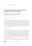
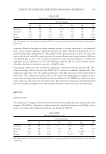
EFFECT OF CYCLOHEXANE AND BENZENE DIESTER ON MELANOGENESIS 177 PREPARATION OF CYCLOHEXANE DIESTER DERIVATIVES (1A–1O) AND BENZENE DIESTER DERIVATIVES (2A–2O) The analogs of cyclohexane diester or benzene diester were synthesized by esterifying fi ve types of acyl chlorides with six types of diol (i.e., 1,2-cyclohexanediol, 1,3-cyclohexane- diol, 1,4-cyclohexanediol, 1,2-benzenediol, 1,3-benzenediol, and 1,4-benzenediol). Ten millimoles of diol was dissolved in 50.0 ml THF, with 12 mmol TEA added slowly. The mixture was stirred for 20 min at room temperature. Then, 12 mmol acryl chloride (butyl chloride, hexanoyl chloride, octanoyl chloride, decanoyl chloride, and 2-ethylhexanoyl chloride) was added and stirred for 6 h at room temperature. Upon completion, the reac- tion mixture was added to water and extracted with ethyl acetate. The extract was washed with brine, dried over MgSO4, and concentrated under vacuum. The residue was purifi ed by fl ash column chromatography on a silica gel. Cyclohexane diester derivatives were prepared using the aforementioned general procedure. MUSHROOM TYROSINASE INHIBITION ASSAY The inhibitory effect of the test compounds against mushroom tyrosinase was examined using the modifi ed method of Masamoto et al. Mushroom tyrosinase (EC 1.14.18.1) used Table I Chemical Structures of Cyclohexene Diester Derivatives (1a–1o) and Benzene Diester Derivatives (2a–2o) Compounds R n Compounds R n 1a–1o 2a–2o 1a –H 1 2a –H 1 1b –H 3 2b –H 3 1c –H 5 2c –H 5 1d –H 7 2d –H 7 1e –CH2CH3 3 2e –CH2CH3 3 1f –H 1 2f –H 1 1g –H 3 2g –H 3 1h –H 5 2h –H 5 1i –H 7 2i –H 7 1j –CH2CH3 3 2j –CH2CH3 3 1k –H 1 2k –H 1 1l –H 3 2l –H 3 1m –H 5 2m –H 5 1n –H 7 2n –H 7 1o –CH2CH3 3 2o –CH2CH3 3

JOURNAL OF COSMETIC SCIENCE 178 for the assay was purchased from Sigma-Aldrich. All test compounds dissolved in DMSO to 500 μM. Two hundred and twenty microliters of 0.1 M phosphate buffer (pH 6.5), 20 μl of sample solution, and 20 μl of mushroom tyrosinase (2000 units/ml in the same buffer) were mixed in a 96-well microplate, and the mixture was preincubated at 37°C for 5 min. Then, 40 μl of 1.5 mM L-tyrosine was added. After incubation at 37°C for 10 min, the absorbance was determined at 490 nm using an ELISA plate reader. % Inhibition = {(A − b)/A} × 100, where A = OD at 490 nm without sample and B = OD at 490 nm with sample. CELL LINES AND CELL CULTURE The mouse melanoma cell line, B16F10, was obtained from Korean Cell Line Bank (KCLB, Seoul, Korea). The cells were cultured in DMEM containing 10% FBS, 50 μg/ml penicillin, and streptomycin at 37°C in the humidifi ed atmosphere of 5% CO2. CYTOTOXICITY TEST Cell viability was determined with a modifi ed version of the method published by Tsukahara et al. (14) using reduction by thiazolyl blue tetrazolium bromide (MTT). B16F10 cells were seeded in DMEM with 10% FBS in separate 96-well plates at the density of 1 × 10⁴ cells/well before being incubated for 24 h. The test compounds were then added to separate wells, and the cells were incubated for another 24 h, after which 100 μl MTT (300 μg/ml) was added to each well. The cells were incubated for 4 h, and 150 μl DMSO was added to dissolve the formazan crystals. The absorbance of the formazan complexes was measured at 540 nm using an ELISA plate reader. IN-VITRO MELANIN SYNTHESIS INHIBITION ASSAY The melanin content of cultured B16F10 cells was determined using a published proce- dure (7) with slight modifi cations. The cells were seeded into six-well plates at the den- sity of 3 × 10⁵ cells/well and incubated for 48 h. Each test sample was then added to the cells, which were subsequently incubated for an additional 24 h. The cells were collected by incubation in trypsin-EDTA followed by centrifugation. Cell pellets were dried, dis- solved in 1 N NaOH, and boiled for 10 min. After cooling, the absorbance was measured at 400 nm using an ELISA plate reader. The absorbance values of a series of known con- centrations of pure melanin were used to construct a calibration curve to determine the amount of melanin produced by the cells. WESTERN BLOTTING B16F10 cell lysates were separated by SDS-PAGE (10% polyacrylamide gels) and transferred to polyvinylidene fl uoride (PVDF). The membranes were then probed with tyrosinase, TRP- 1, TRP-2, and tyrosinase antibodies. Briefl y, the cultured B16F10 cells were washed with phosphate-buffered saline and incubated with a RIPA lysis buffer (1% Nonidet P-40, 1%
Purchased for the exclusive use of nofirst nolast (unknown)
From: SCC Media Library & Resource Center (library.scconline.org)