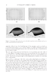
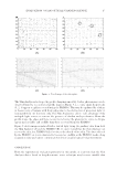
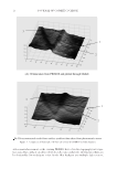
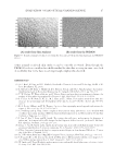
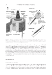
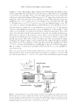
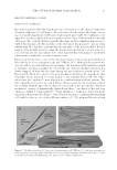
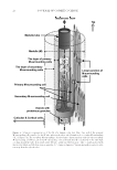
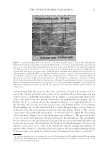
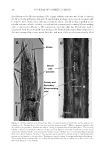
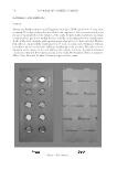
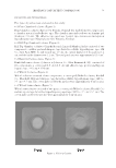
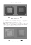
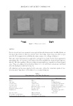
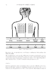
J. Cosmet. Sci., 69, 19–33 ( January/February 2018) 19 New Aspects of the Structure of Human Scalp Hair-II: Tubular Structure and Material Flow Property of the Medulla ASAO YAMAUCHI and KIYOSHI YAMAUCHI, Biomaterials Research Division, Osaka Research Institute of Industrial Science and Technology, 1-6-50 Morinomiya, Joto-ku, Osaka 536-8553, Japan (A.Y.) and Keratin Materials Research Laboratory, 1-5-3-308 Kitakutsukake, Nishikyo-ku, Kyoto 610-1101, Japan (K.Y.) Accepted for publication September 1, 2017. Synopsis Asian scalp hair fi bers were made thin by treatment with papain or sliced along the longitudinal axis or randomly cut by mechanical means. Optical microscopic observations of the resulting specimens indicated that (i) the medulla (M) consisted of two types of the M-surrounding cells which were linearly linked one another to form a tubular structure running through the fi ber and (ii) the drum-shaped vesicles containing small proteinous granules were neatly or sparsely stored within the tube. On the other hand, H+ and OH- ions were able to move spontaneously from one end to another through the M tube. Large molecules such as an anthocyanin dye (from purple sweet potato) were also capable of fl owing through the M tube, especially rapidly when DC voltage was applied between the two ends of the hair fi ber. The possible function of the M is briefl y discussed in conjunction with the tubular structure and the material fl ow property. INTRODUCTION Previously we reported the new structural model of an Asian scalp hair fi ber, especially focusing on the morphology and the spatial arrangement of the cortical and cuticular cells (1) see Figure 1. Although a lot of studies have been carried out over the last several decades using the optical and electron microscopes (2–10), our experimental method, which was characterized by random scission of the fi bers, led to many revisions of the traditional hair structural model (7,11). The present investigation concerns the medulla, which exists in the middle of the fi ber and has two aims. The fi rst, is to obtain the detail structure of the medulla. Optical micros- copy was fully used in the study in other words, whereas electron microscopy works in vacuo and provides only a black-and-white photograph of the dried substance, the optical Address all correspondence to Asao Yamauchi at yamauchi@omtri.or.jp.
Purchased for the exclusive use of nofirst nolast (unknown)
From: SCC Media Library & Resource Center (library.scconline.org)