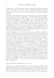
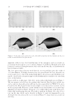
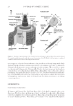
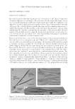
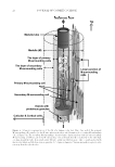
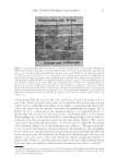
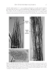
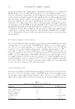
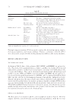
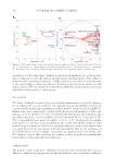
JOURNAL OF COSMETIC SCIENCE 20 Figure 1. Sche matic representation of the overall structure of a human scalp hair fi ber. The previous model (1) is partly modifi ed to match with the present observations of the M. Each of the hair parts is explained elsewhere in the text. The unit for the length is micrometer. microscopy not only suits for wet substances but provides a see-through image with a high depth of fi eld in semi-micro and micrometer levels, often making it possible to distinguish chromatically between two or more substances in the specimen. The second, is to exam- ine the material fl ow property of the medulla. To date, the hair component has long been considered just a loosely packed assembly of the cellular disks connected in series and not performing any signifi cant function in hair (9,11–16). The more detailed structural anal- ysis of the medulla and its function, if any, may be very useful to evaluate the interaction of human hair with various kinds of chemical and biochemical substances including cosmetic products. EXPERIMENTAL HAIR SAMPLES AND REAGENTS A Japanese girl donated her black hair fi bers (jf8) cf. the Arabic numeral refers to the donor age. The fi bers were cut from more than 1 cm from the scalp surface. Black hairs (cf16) were similarly collected from a girl of the Miao ethnic group living in the mountains of China’s Yunnan province. White hairs, jf65 and jm67, were furnished from Japanese

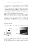
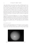
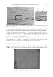
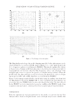
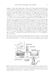
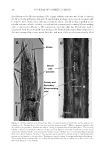
STRUCTURE OF HUMAN SCALP HAIR−II 21 female and male, respectively. Although it is not mentioned herein, various other persons also donated their scalp hairs to the present study. All of the fi bers were straight, neither being stained with dyes nor subjected to any permanent setting processes. The fi bers were succes- sively washed with a 1.5% (w/w) aqueous solution of sodium dodecyl sulfate (SDS), deionized water, and 70% (v/v) ethanol and then stored at 4°C in a sealed plastic container. The reagents including an anthocyanin dye (from purple sweet potato a mixture of cyanidin acylglycoside and peonidin acylglycoside Kiriyachemi, Osaka, Japan) and purifi ed papain powder from Carica papaya (Wako Pure Chemical Industries, Osaka, Japan) were commercially available. MICROSCOPE OBSERVATION The biological microscope, Olympus, Tokyo, Japan model Vanox (AH), was used mainly for bright fi eld, phase contrast, and polarized light observations. The inverted microscope and the stereomicroscope, Olympus model CK30 and X-Tr, respectively, were modifi ed so as to place the observation chambers and the electrode cells (vide infra) on the micro- scopes. The microscopes were equipped with a digital camera, which was automatically controlled by a desktop computer to optimize for lighting, ISO levels, and focusing mea- sures. The images in JPEG and RAW formats were developed by means of Lightroom ver. 3 and Photoshop Elements ver. 9 software (Adobe Systems Inc., San Jose, CA). Image en- hancement included color level correction, noise reduction, and contrast and brightness adjustments the purpose of the enhancement was to make the image nearly identical to that seen actually by the observers. In the case of thick specimens, the images taken at various depths of fi eld were merged into a single deep focus picture by the use of the stacking software, Combine ZP (A. Hadley, Sheffi eld, United Kingdom) (17). SPECIMEN PREPARATION FOR STRUCTURAL ANALYSIS OF MEDULLA Enzymatic thinning of hair fi bers using the papain (a representative procedure). Several strands of the hair (jf8, about 15 mm in length) were warmed in pH 7/0.067 M phosphate buffer solution (PBS) at ambient temperature for 24 h. After washing briefl y with water and drying at ambient temperature, the fi bers, usually 1–3 strands, were placed parallel on a glass plate (22 × 22 × 0.17 mm) and both ends of each fi ber were glued to the plate with a rapid type–epoxy resin see Figure 2(A). Next, the glass plate having the fi bers was fi xed by the use of the epoxy resin on the window frame (20 × 20 mm) which had been made in the bottom of a plastic culture dish (diameter, 33 mm depth, 10 mm). After washing with PBS overnight at ambient temperature, the papain solution (20 units in 3 mL of PBS) containing its activator of SDS (12 mg) and 2-mercaptoethanol (ME) (65 μL) was put into the dish, and the digestion of the fi bers was allowed to proceed at 30°C. The inverted microscope was used to observe the morphological change of the fi bers. Random mechanical cutting of enzymatically thinned hair fi bers (a representative procedure). Sev- eral strands of the white fi bers (jf65 about 15 mm in length) were treated by dipping in PBS at ambient temperature for 24 h, and then warmed in the PBS solution (3 mL) of papain (20 units), SDS (12 mg), and ME (65 μL) at 30°C with occasional gentle shaking. After about 3 h, a new crop (10 units) of the enzyme was added to the reaction mixture. During incubation, one strand of the fi bers was periodically sucked into water (1 mL)
Purchased for the exclusive use of nofirst nolast (unknown)
From: SCC Media Library & Resource Center (library.scconline.org)