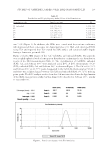
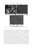
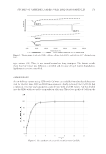
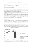
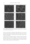
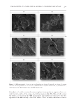
JOURNAL OF COSMETIC SCIENCE 184 reticulated by LOXL1. Indeed, we have shown that LOXL1 is downregulated under UV radiation. Moreover, LOXL1 has been shown to be degraded by skin elastases. Abnormal and nonfunctional fi brils appear. Furthermore, elafi n is a serine protease inhibitor that is mostly produced by epithelial cells. In the skin, keratinocytes are the main source of this molecule. Although elafi n is not detectable in normal skin, it is abundantly secreted in psoriasis and other infl amma- tory skin disorders (10). Elafi n acts in a varied way on the cutaneous immune homeosta- sis. Elafi n does not only exert an antiprotease effect but also an immunomodulatory and antiproliferative one (11). In solar elastosis, fi broblasts also express elafi n, which binds to elastin. The elafi n–elastin complex limits the elastolytic regular process, leading to the accumulation of disintegrated abnormal elastic fi bers and aggregates. The UV-induced elafi n prevents the proteases to degrade the abnormal fi bers (9). By inhibiting the synthe- sis of elafi n, our specifi c Hamamelis extract gives access back to proteases, to degrade the abnormal fi bers. Therefore, the specifi c Hamamelis extract acts at two levels: (i) it makes more functional elastin fi bers by increasing the elastin–cross-linked enzyme LOXL1 and (ii) prevents the formation of the nonfunctional elastin fi bers by inhibiting elafi n, the peptidase inhibitor. The in vitro results are confi rmed with the clinical data which show a decrease in wrinkles and an improvement in the fi rmness of the skin. CONCLUSION Complementarily to UV fi lters’ sun protection, we developed an active ingredient able to give the skin its own power to fi ght against the damage caused by the sun and to repair the already existing ones. Thanks to our knowledge on elastic fi bers, we discovered a process triggered in response to UV light, which upsets the balance of these fi bers: too much elastin and too little LOXL1 enzyme to assemble them into functional elastic fi bers. This imbalance leads to the accumulation of nonfunctional elastin, which groups together in aggregates and cannot be naturally removed. In addition to this imbalance, the fi bro- blast synthesizes elafi n, known to be a marker of elastotic aggregates. This protein crys- tallizes the elastin fi bers and emphasizes the formation of aggregates, which cannot be naturally eliminated by the skin. We have designed the fi rst active ingredient based on Hamamelis virginiana leaf extract (witch hazel) that corrects the damage caused by solar elastosis and that acts as a powerful shield against photoageing by acting on these two phenomena: the imbalance between LOXL1 and elastin, and the overexpression of elafi n. The active ingredient effi cacy is proven: lines are decreased and the skin recovers its fi rmness. REFERENCES (1) D. C. Calderone and N. A. Fenske, The clinical spectrum of actinic elastosis, J. Am. Acad. Dermatol., 32, 1016–1024 (1995). (2) M. J. Koehler, S. Zimmermann, S. Springer, P. Elsner, K. König, and M. Kaatz, Keratinocyte mor- phology of human skin evaluated by in vivo multiphoton laser tomography, Skin Res. Technol., 17, 17479–17486 (2011). (3) J. Uitto, The role of elastin and collagen in cutaneous ageing: intrinsic ageing versus photoexposure, J. Drugs Dermatol. 7, s12–s16 (2008).

UNDERSTANDING SOLAR SKIN ELASTOSIS 185 (4) J. Uitto, M. J. Fazio, and D. R. Olsen, Molecular mechanisms of cutaneous ageing. Age-associated connective tissue alterations in the dermis, J. Am. Acad. Dermatol., 21, 614–622 (1989). (5) J. M. Davidson, S. Shibahara, C. Boyd, M. L. Mason, P. Tolstoshev, and R. G. Crystal, Elastin mRNA levels during foetal development of sheep nuchal ligament and lung. Hybridization to complementary and cloned DNA, Biochem. J., 220, 653–663 (1984). (6) R. P. Mecham, T. Broekelmann, E. C. Davis, M. A. Gibson, and P. Brown Augsburger, Elastic fi bre assembly: macromolecular interactions, Ciba Found. Symp., 192, 172–181 (1995). (7) G. C. Sephel, A. Buckley, and J. M. Davidson, Developmental initiation of elastin gene expression by human fetal skin fi broblasts, J. Invest. Dermatol., 88, 732–735 (1987). (8) R. Pfundt, I. Van Vlijmen-Willems, M. Bergers, M. Wingens, W. Cloin, and J. Schalkwijk, In situ demonstration of phosphorylated c-jun and p38 MAP kinase in epidermal keratinocytes following ultraviolet B irradiation of human skin, J. Pathol., 193, 248–255 (2001). (9) J. Muto, K. Kuroda, H. Wachi, S. Hirose, and S. Tajima, Accumulation of elafi n in actinic elastosis of sun damaged skin: elafi n binds to elastin and prevents elastolytic degradation, J. Invest. Dermatol., 127, 1358–1366 (2007). (10) J. Muto, N. Fujimoto, K. Ono, T. Kobayashi, K. R. Chen, S. Suzuki, H. Wachi, and S. Tajima, Deposition of elafi n in the involved vascular wall of neutrophil-mediated cutaneous vasculitis, J. Eur. Acad. Derma- tol. Venereol., 30, 1544–1549 (2016). (11) T. Verrier, B. Solhonne, J. M. Sallenave, and I. Garcia-Verdugo, The WAP protein trappin-2/elafi n: a handyman in the regulation of infl ammatory and immune responses. Int. J. Biochem. Cell Biol., 44, 1377–1380 (2012).
Purchased for the exclusive use of nofirst nolast (unknown)
From: SCC Media Library & Resource Center (library.scconline.org)