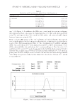
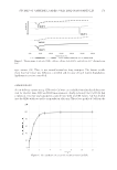
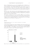
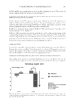
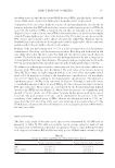
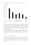
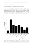
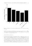
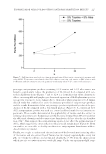
JOURNAL OF COSMETIC SCIENCE 206 collect stratum corneum from the volar forearm of volunteers. Products were sprayed onto the isolated corneocytes from an aerosol placed about 12 inches away from the stratum corneum. The products were left to air-dry for a minimum of 20 min before coating with conductive metals and placing into the scanning electron microscopy (SEM) chamber. SEM PROTOCOL Treated D-Squame discs were placed on 25 mm OD Pelco Tabs (Ted Pella, Inc., Redding, CA). The pin stubs were placed in a sputter coater (Leica EM ACE600 Leica Microsys- tems, Wetzler, Germany) and coated for 60 s with gold/palladium, resulting in a 10-nm thick layer. The stubs were attached to a multisample mount and placed into the SEM (Hitachi SU-5000, Tokyo, Japan), which has variable pressure and fi eld emission scan- ning capabilities. For the analysis of the coated stratum corneum layers, we used high vacuum mode ( 1 × 10⁻³ Pa) and collected photomicrographs starting at ×300 and extending to higher magnifi cations to elucidate details associated with the sunscreen ap- plication. Most images were collected using a secondary electron detector, but a backscatter detector was also used for samples that exhibited poor contrast properties. 3D images were also captured by SEM. RESULTS In this study, we sought to determine key properties of sunscreen preparations applied to skin. Using evaporimetry in conjunction with a Franz diffusion cell apparatus and ex vivo skin, vapor fl ux data were generated for the sunscreens, allowing for the determination of the water permeability of the sunscreen fi lms. Employing SEM, we developed a novel methodology for determining the morphological fi lm properties when sunscreens are ap- plied to skin. The utility of the technique, aimed at discerning the microscopic fi lm properties, lies in the use of layers of corneocyte cells collected by tape stripping. In this manner, the substrate has the same surface properties as in vivo skin. EVAPORIMETER STUDIES Four formulations were tested in this study, namely, Formulations C, D, E, and F. Because Formulations A and B consist mostly of alcohol, their water permeability was not tested. Instead, water and sunfl ower seed oil were used as negative and positive controls and yielded a cumulative evaporation of 88.0 × 10³ and 12.6 × 10³ g m-2 h-1, respectively. Both the positive and negative controls were signifi cantly different from all treatments and from each other. The data generated for the four formulations studied are displayed in Figure 1. Formulations C, D, E, and F yielded cumulative evaporations of 53.4, 37.7, 46.0, and 36.6 × 10³ g m-2 h-1, respectively. Only Formulation C was signifi cantly differ- ent (when comparing Formulations C, D, E, and F), in which case there was a greater rate of evaporation. Formulations D, E, and F, which contained VA/butyl maleate/isobornyl acrylate copolymer and its combination with acrylates/dimethicone copolymer or hydroxypropyl cellulose,

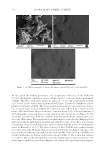
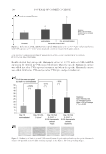
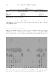
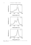
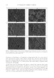
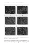
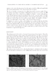
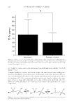
FILM PROPERTIES OF POLYMERS USED IN ANHYDROUS SUNSCREEN FORMULATIONS 207 formed signifi cantly less permeable fi lms than Formulation C, which did not contain any polymer. Cumulative evaporimeter data indicated that the sunscreen phase had the high- est reduction in cumulative evaporation rate, specifi cally 39.3%/h, followed by the addi- tion of a fi lm former to the spray which reached an additional reduction of 17.9%/h in the best case. These fi ndings are quite reasonable, as sunscreen fi lter concentrations are typically much higher (typically 20–30% w/w) than polymer concentrations (typically 1–3% w/w) in the fi nal formulation. The data from the evaporimeter studies confi rmed that the sunscreen fi lters play a more important role in water vapor transmission than the polymers added to the formulation. This fi nding seems to correlate with previously presented data (6). It is also important to note that the addition of polymers to the sunscreen formulations increased its in vitro water resistance. MICROSCOPIC EVALUATION OF FILMS ON SKIN To better understand the deposition characteristics of the sunscreen systems on skin, we used SEM to carefully monitor the deposition behavior of sunscreen fi lms and to elucidate the architectural role played by several polymeric systems in the sunscreens. Figures 2 and 3 contain micrographs of corneocytes treated with all of the studied formulations at ×300 (Figure 2) and ×500 or ×1,000 (Figure 3) magnifi cations, respectively. Figure 1. Evaporation data obtained from sunscreen fi lms applied to ex vivo porcine skin in a Franz diffusion cell apparatus.
Purchased for the exclusive use of nofirst nolast (unknown)
From: SCC Media Library & Resource Center (library.scconline.org)