JOURNAL OF COSMETIC SCIENCE 216 the ability of the cell’s mitochondria to reduce the yellow MTT to a purple formazan (crystals) end product. The reaction was terminated by the removal of the MTT solution. Addition of 1.0 ml of 0.04 M HCl in isopropanol to each well was used to dissolve the intracellular MTT formazan crystals. The contents of each well were mixed gently in an orbital shaker at room temperature for 1 h and the absorbency at 570 nm was measured by an enzyme-linked immunosorbent assay (ELISA) microplate reader (SpectraMax M5 Multi-Mode Microplate Readers Molecular Devices, San Jose, CA). Cell viability data obtained from the MTT assay were used to normalize IL-1α and IL-8 levels. CYTOKINE RELEASE MEASUREMENTS To assess the irritancy potential of aluminum-containing solutions and formulations, in vitro experiments were conducted using MatTek EpiDerm tissue model to measure the release of infl ammatory cytokines. In vitro skin samples were treated topically with 30 μl of solutions or products for 1 h in an incubator at 37°C and 5% CO2. Following the in- cubation, skin samples were washed with PBS and placed in culture medium, provided by MatTek Corporation, and continued to incubate for 24 h. After 24 h, cell culture media were collected for IL-1α release assay. IL-1α levels were analyzed by the ELISA assay kit (R&D Systems, Minneapolis, MN). IL-8 levels were analyzed using immunoas- say multiplex kit (Millipore, Billerica, MA) on a Luminex X200 (Luminex Corporation, Austin, TX). PERCUTANEOUS ABSORPTION OF TAURINE Percutaneous permeation of taurine was evaluated in vitro by using human keratinocyte skin model, EpiDerm 200-X. A standard permeation device, MatTek Permeation Device was used to approximate the permeability of taurine through the skin. 5% taurine solution with or without 5% aloe extract (w/w) was applied topically to EpiDerm tissue. After set time points the EpiDerm tissue, donor and receiver solutions were collected and ana- lyzed for taurine concentration. Receiver solution was 5.0 ml PBS solution and stored at -20°C. DETERMINATION OF CELLULAR TAURINE CONCENTRATION High-pressure liquid chromatography (HPLC) was used to determine cellular taurine concentration after treating MatTek EpiDerm tissue with a 5% (w/w) taurine solution or 5% (w/w) taurine and aloe extract solution. Before analysis, tissue samples were lysed using an ultrasonic tissue lyser, Qiagen Retsch MM300 (Retsch Inc, Newtown, PA) in 500 μl of 5× Extraction Buffer (Enzo Life Sciences Inc., Farmingdale, NY). Tissue lysates were pooled together from the same treatment groups (n = 2) to ensure the adequate vol- ume needed to perform the taurine analysis. Taurine does not absorb UV/Vis radiation adequately thus, a pre-column derivatization reaction is necessary to allow for detection by HPLC. 4-fl uoro-7-nitrobenzofurazan (NBD-F) was used as a fl uorescent reagent to produce derivatives of primary and secondary amines. NBD-taurine derivative has a

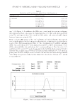
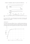
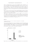
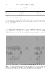
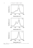
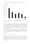
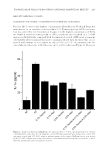
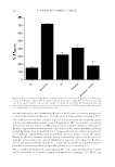
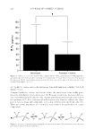
TAURINE/ALOE VERA FOR BOOSTING ANTI-SKIN IRRITATION EFFECTS 217 maximum UV/Vis intensity at 470 nm and has high specifi city. Precolumn derivatization was performed as described in Pencheva et al. (18). Separation was performed using an isocratic elution with TBAB/phosphate (0.01 M/0.08 M) buffer: acetonitrile (70:30, v/v) mobile phase with a fl ow rate of 1 ml/m at a 7 m runtime. Chromatographic separations were performed on a Zorbax C18 (4.6 × 150 mm, 5 μ) column. Data were collected with an Agilent 1200 G1310A Isocratic pump, G1329A Autosampler, G1314B Variable Wavelength Detector. Analysis was performed with Agilent Chemstation software (Agilent Technologies, Inc., Wilmington, DE). Concentration of taurine was calculated based on peak area by using a regression curve constructed from a six-point (10.43–208.8 ppm) taurine calibration curve. Standard solutions were prepared from a stock taurine solution and regression curve had a correlation coeffi cient of 0.991. SIZE-EXCLUSION CHROMATOGRAPHY (SEC–HPLC) To ensure taurine and aloe extract did not affect the high effi cacy of AlCl3 antiperspi- rant salt, the size distribution of aluminum salt in aqueous solutions was monitored by SEC or SEC–HPLC. Retention times for each of the peaks may vary depending on ex- perimental conditions but they remain relative to each other. Water®600 analytical pump and controller, Rheodyne®7725I injector using a Protein-Pak® 125 (Waters) column, and Waters 2414 Refractive Index Detector were used to collect SEC data. The mobile phase was a 5.56 mM nitric acid solution, pH of 2.3 (with KNO3), with a fl ow rate of 1.0 ml/min. Analysis was conducted using Waters® Empower software (Waters Corporation, Milford, MA). Peak distribution for aluminum salt in our prototype AlCl3 formulation was observed to be similar to a 1.2% AlCl3 in DI water solution, which suggests that taurine and aloe extract would not affect the high effi cacy of AlCl3 salt. When the extraction of AlCl3 from our prototype product for SEC was conducted, the concentration of AlCl3 is reduced. However, the elution time for the aluminum peaks are the same, which indicates that the aluminum chloride is intact. If taurine and aloe extract hydrolyze aluminum chloride to large, insoluble aluminum hydroxide species, we would expect multiple peaks to elute before the aluminum chloride peak but we only observe one aluminum peak, peak 5, at a retention time of ca. 9.0 min (Figure 1). CLINICAL STUDY DESIGN A human clinical study was conducted to compare the anti-irritating properties of taurine and aloe extract in a 12% AlCl3 antiperspirant prototype product to a 12% com- mercial AlCl3 antiperspirant product. The study enrolled six male and six female subjects (n = 12) and lasted 4 days. Each subject was treated with both products (one on each desig- nated forearm area) in a randomized design. A 5 cm by 5 cm area was marked on both forearms where the prototype product and benchmark product were applied. Dosage for both products was 3 mg/cm2. Products were applied once a day, for four consecutive days in the mornings and were left on the skin overnight. Skin surface samples were collected at least 24 h after the fourth application to analyze for IL-1α levels. Skin surface samples were collected by using the cup-scrubbing method. A hollow glass cylinder (8.5 cm2) was
Purchased for the exclusive use of nofirst nolast (unknown)
From: SCC Media Library & Resource Center (library.scconline.org)