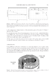
J. Cosmet. Sci., 69, 315–322 (September/October 2018) 315 Bridging the “Dead Hair”—“Live Follicle” Divide in Applied Hair Research MARTA BERTOLINI, NATALIA BOTCHKAREVA, GILL WESTGATE, CHRIS WARD, PAUL CORNWELL, and RALF PAUS, Monasterium Laboratory, Skin and Hair Research Solutions GmbH, Muenster, 48149, Germany (M.B., N.B., C.W.), Centre for Skin Sciences, University of Bradford, Bradford, BD7 1DP England (G.W.), Textile Research Institute, Princeton, New Jersey, 08540, (P.W.), University of Manchester, Manchester, M13 9PT United Kingdom (R.P.), University of Miami, Miami, 33125, Florida (R.P.) Synopsis For many decades, applied hair research has been hampered by an unproductive intellectual and conceptual divide between researchers who are primarily interested in the hair shaft (HS), its structural properties, visual appearance and cosmetic manipulation, and those investigators who are mainly interested in the fascinating miniorgan that cyclically regenerates the HS, the hair follicle (HF). This article attempts to bridge this unproductive divide between the “dead hair” and “live follicle” worlds by summarizing both current key concepts and major open questions on how the HF, namely, the anagen hair bulb and its precortical hair matrix keratinocytes, generate the HS, focusing on selected key signaling pathways. We discuss current theories of hair shape formation and avenues toward impacting on human HS structure. The article closes by delineating which instructive preclinical research assays are needed to ultimately close the experimental gap between HS and HF researchers in a manner that benefi ts consumers. The hair follicle (HF) is a unique miniorgan whose main function is to produce a pig- mented hair shaft (HS) (1–3). The number of HFs in the human body, about 5 million, is determined during embryogenesis when HFs develop as the result of a bidirectional cross-talk between cutaneous ectoderm and mesenchyme (4–6). During fetal life, lanugo HFs generate soft but long fi ne fi laments, which are present during the prenatal period and are usually shed in utero or during the fi rst weeks of life. These fi rst hairs are then replaced by vellus HFs that develop into terminal HFs only in particular body areas. Vellus HFs form short, unmedullated, non- or light-pigmented hair and cover most areas of the body surface. Terminal HFs, which give rise to long, thick, and pigmented hair, are developed in the scalp or after puberty in androgen-dependent body areas, such as the pubic area, underarm, and male beard (2,7). Address all correspondence to Marta Bertolini at m.bertolini@monasteriumlab.com.
Purchased for the exclusive use of nofirst nolast (unknown)
From: SCC Media Library & Resource Center (library.scconline.org)